Click on images to enlarge
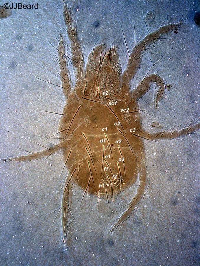
Fig. 1. Eotetranychus pronus adult female - dorsal habitus.
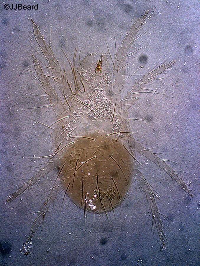
Fig. 2. Eo. pronus adult female - dorsal habitus.
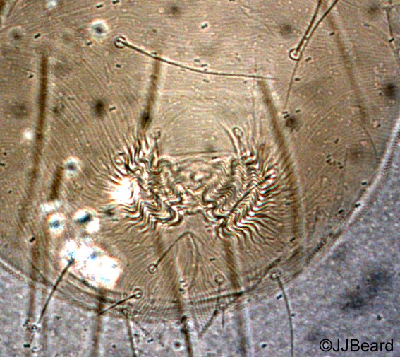
Fig. 3. Eo. pronus adult female - detail of pattern of pregential striae.
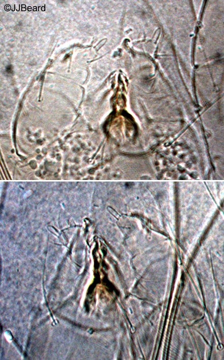
Fig. 4. Eo. pronus adult female - detail of palp, showing elongate spinneret.

Fig. 5. Eo. pronus adult female - detail of tarsus I.

Fig. 6. Eotetranychus pronus adult male - lateral habitus.
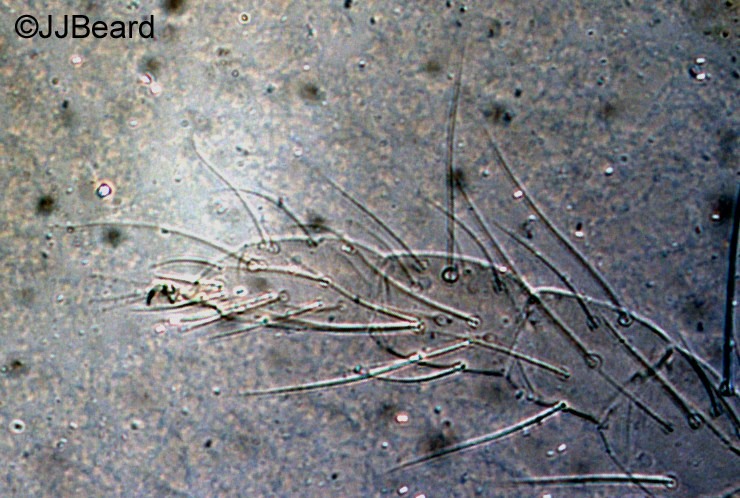
Fig. 7. Eo. pronus adult male - detail of tarsus I.
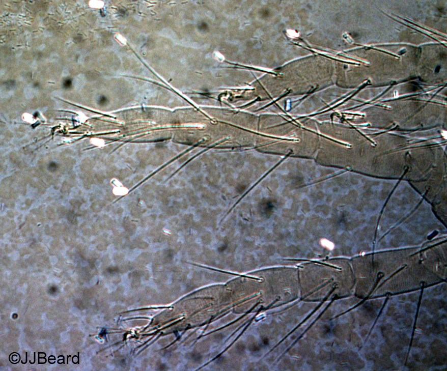
Fig. 8. Eo. pronus adult male - detail of tarsus III & IV.

Fig. 9. Eo. pronus adult male, detail of aedeagus - a. holotype (different focus levels through specimen); b. paratype (different focus levels); c-d. topotypes (with or without associated structures illustrated).
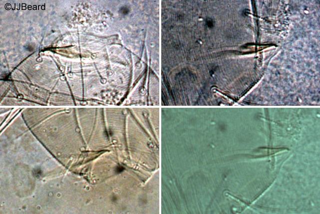
Fig. 10. Eo. pronus adult male - detail of aedeagus.
Material examined
types
Taxonomy
Subfamily Tetranychinae
Tribe Tetranychini
Distribution
*Australia: south eastern Queensland
Taxonomy Changes
None
Diagnosis
Female (Figs 1,2)
- empodia I-IV = proximoventral hairs only, no dorsal spur/claw
- peritreme ending in slightly expanded oblong simple bulb
- dorsal opisthosomal striae transverse
- dorsal setae conspicuously pubescent
- lobes on striae small, semi-circular, widely spaced
- ventral striae mostly without lobes
- pregenital striae transverse (Fig. 3)
- palp with spinneret elongate, 3-4 times as long as wide (Fig. 4)
- tarsus I with sockets of four-five tactile setae and one solenidion proximal to the socket of the proximal duplex seta (Fig. 5)
- tarsus II with sockets of two-three tactile setae and one solenidion proximal to the socket of the duplex seta
- tibia I-IV 10(1+0), 8, 6, 7
- yellow-green with several dark spots laterally along body, including a prominent dark spot caudally on each side of the body
- eggs are globular with long dorsal stipe, pearly white, changing to amber when eye spots of the larva appear through the cuticle; laid singley in the webbing of the colony, not fixed to the leaf surface
Male (Fig. 6) as per female plus:
- empodium I claw-like, with some associated hairs (Fig. 7)
- empodia II-IV as in female (Fig. 8)
- peritreme ending in slightly expanded simple bulb
- spinneret on palp tiny, conical
- tarsus I with sockets of four tactile and two-three solenidia proximal to the socket of the proximal duplex seta
- tarsus II with sockets of three tactile setae and one-two solenidion proximal to the socket of the duplex seta
- tibia I-IV 13(4+0), 8, 6, 6-7
- aedeagus almost straight, directed slighty ventrally, tapering towards tip, then abruptly bent ventrally and ending in small anvil-shaped truncate knob; anterior projection short triangular, posterior projection slightly longer, sharp; ventral margin of knob flat; ventral margin of shaft is almost straight, dorsal margin straight but at 30°angle to ventral margin (Figs 9, 10)
Hosts
*Ficus coronata, Ficus spp. (Moraceae), Rubus rosifolius (Rosaceae)
Similar Taxa
Eotetranychus cernuus Baker & Pritchard, E. perplexus (McGregor), E. edi Meyer, E. mastichi De Leon, E. friedmanni Gutierrez, and E. vaughni Baker & Pritchard all have a similarly shaped aedeagus, with the most similar being E. cernuus.
Eotetranychus cernuus and E. pronus are separated based on leg chaetotaxy, spinneret shape, host plant and country of origin: E. cernuus male tibia II with 7 tactile setae, E. pronus male tibia II with 8; E.c. female with 4 tactile setae proximal to duplex setae on tarsus I, E.p. with 5 tactile setae proximal; E.c. female spinneret twice as long as wide, E.p. female spinneret 3 times as long as wide; E.c. host plant = Anona chrysophylla, E.p. host plant = Ficus coronata; E.c. found in Congo, E.p. found in Queensland, Australia.
Biology
Colonies of E. pronus occur on the thickly haired ventral surface of the host plant's leaves. Little to moderate amounts of webbing is produced and mite feeding causes typical spider mite damage to host leaves, that of light speckling.
References
*Davis, J.J. (1969d) Studies of Queensland Tetranychidae (Acarina: Prostigmata) 6. A new genus and five new species of spider mites from native plants. Memoirs of the Queensland Museum 15: 165-183
Copyright © 2018. All rights reserved.