Click on images to enlarge
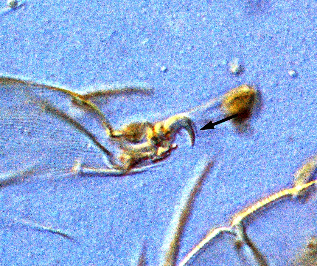
Fig. 1. Schizotetranychus baltazari adult female paratype - detail of claw (arrow indicates a dorsal "hair").
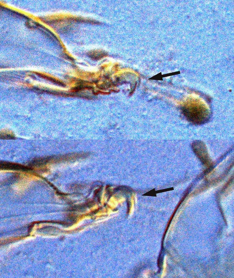
Fig. 2. Schizotetranychus baltazari adult female paratype - detail of claws III, IV (arrow indicates a dorsal "hair").

Fig. 3. Schizotetranychus baltazari adult female paratype - detail of the pattern of pregenital striae.
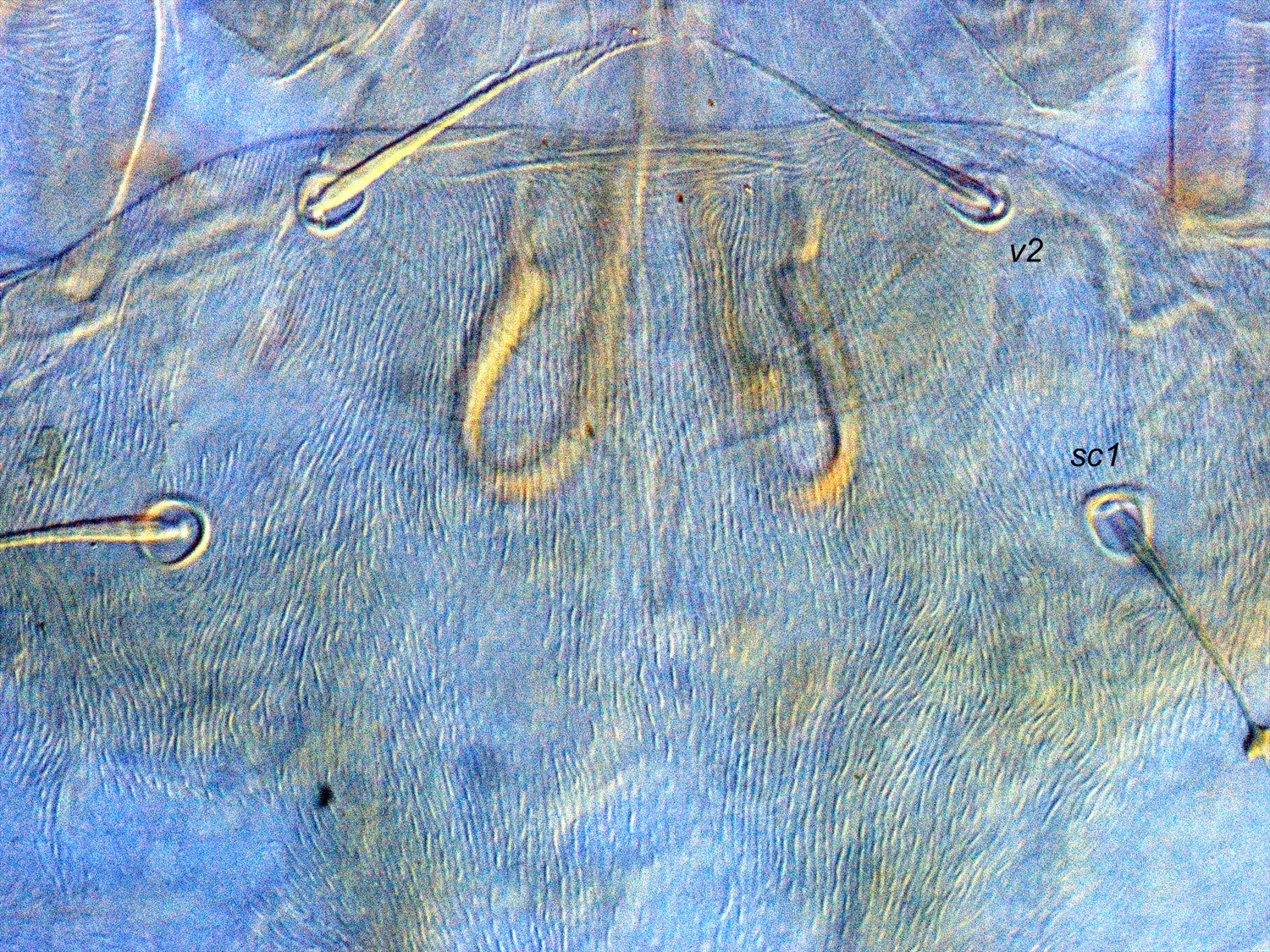
Fig. 4. Schizotetranychus baltazari adult female paratype - detail of the pattern of prodorsal striae.

Fig. 5. Schizotetranychus baltazari adult female paratype - detail of the pattern of prodorsal striae.
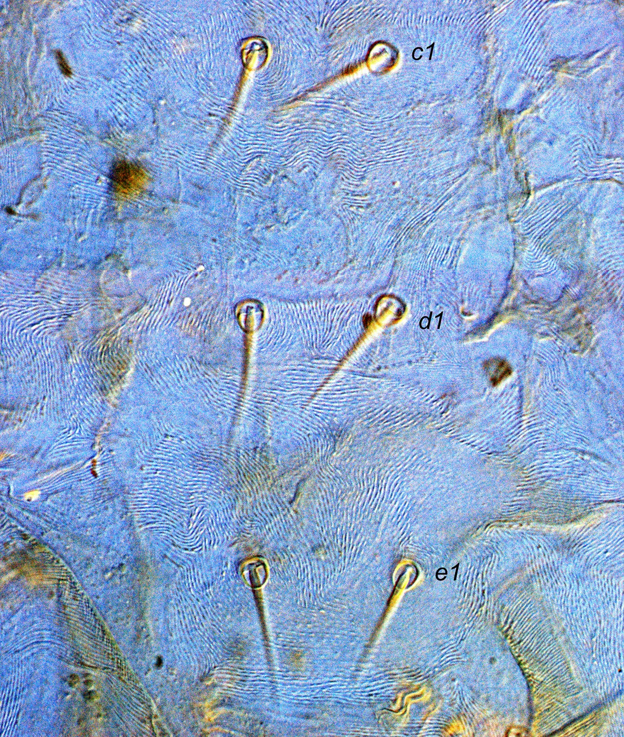
Fig. 6. Schizotetranychus baltazari adult female paratype - detail of the pattern of dorsal striae from setae c1-e1.
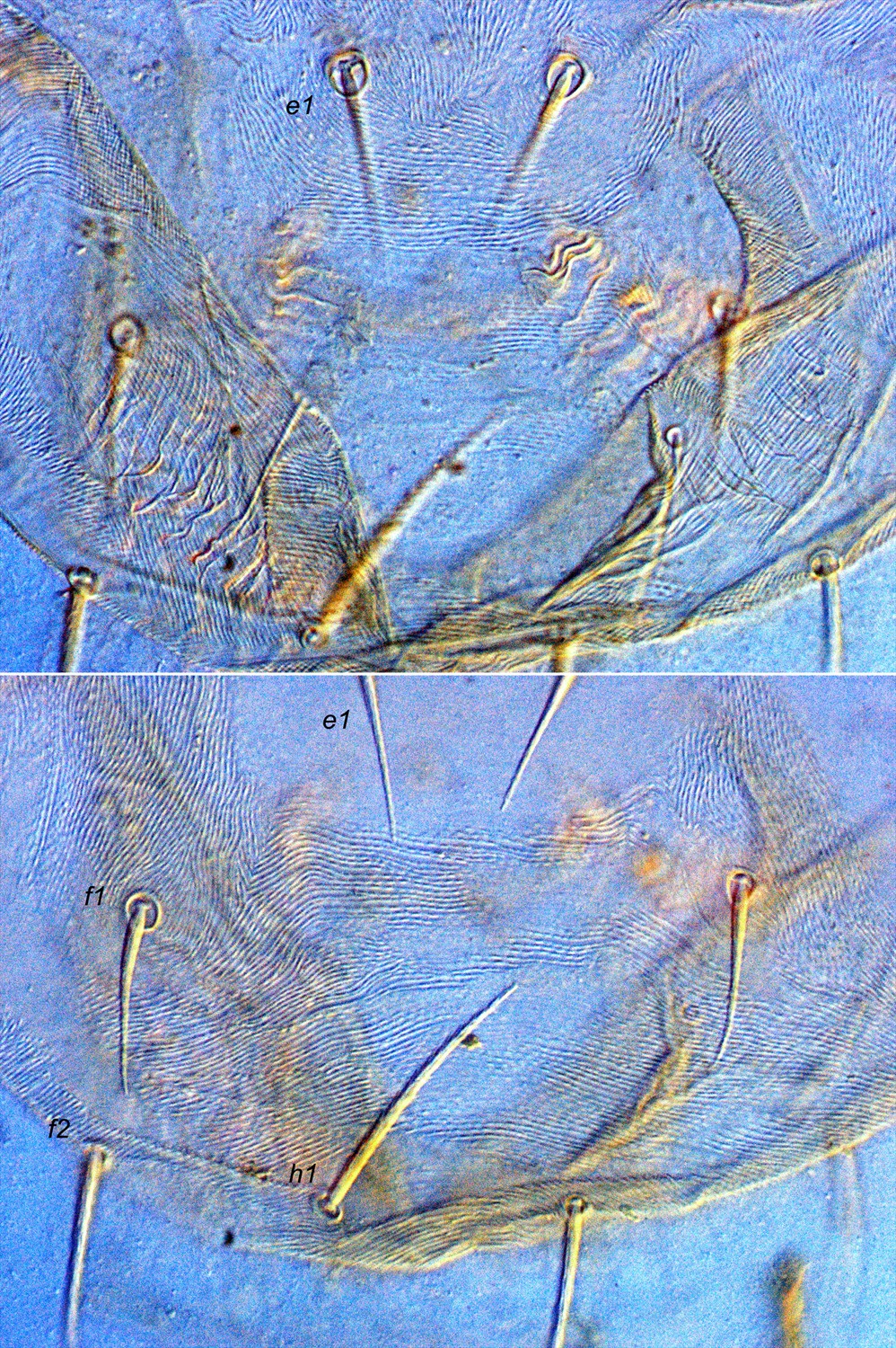
Fig. 7. Schizotetranychus baltazari adult female paratype - detail of the pattern of dorsal striae from setae e1-h1.
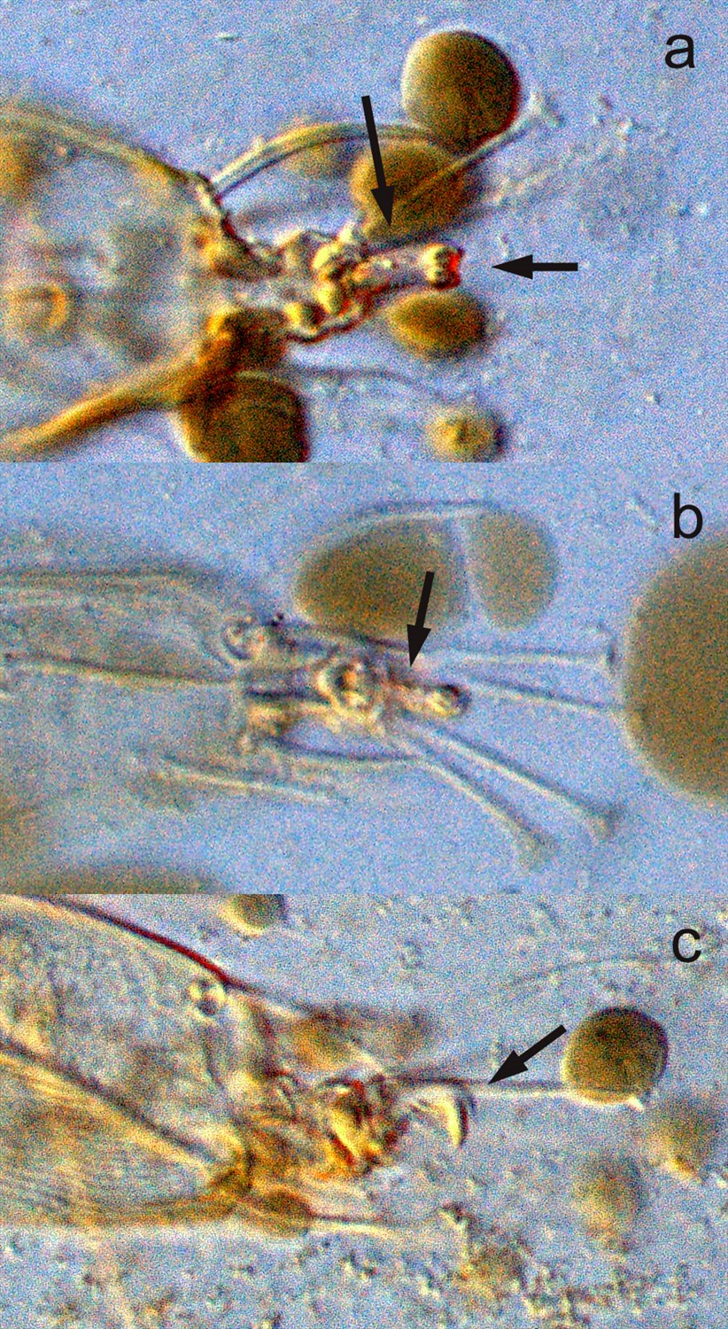
Fig. 8. Schizotetranychus baltazari adult male paratype - detail of claws I, II, III - a. ventral aspect showing basal undivided stalk, with minute proximoventral hairs (indicated by arrow - dots), and distal fork (indicated by arrow); b. ventral aspect with minute proximoventral hairs (indicated by arrow - dots); c. lateral aspect with thin dorsal hair indicated by arrow.
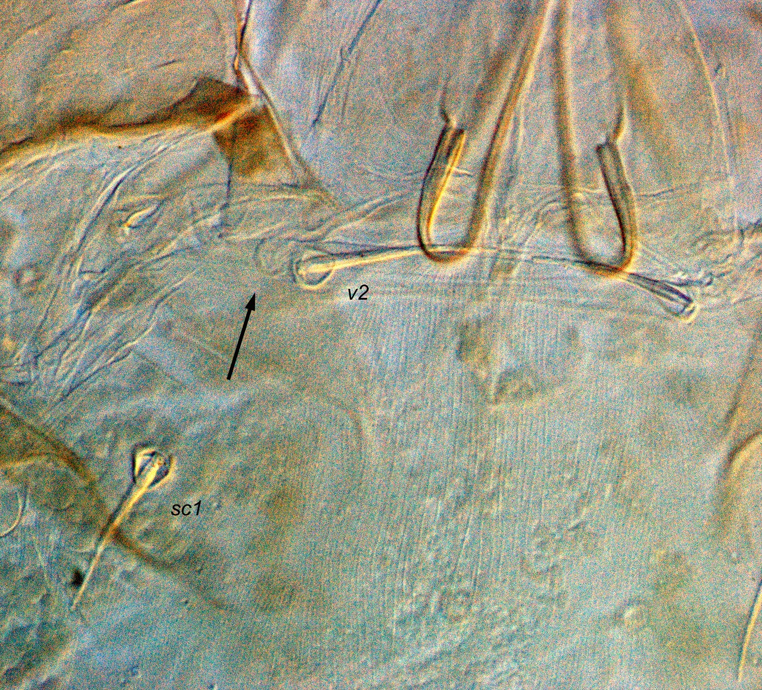
Fig. 9. Schizotetranychus baltazari adult male holotype - detail of peritreme.
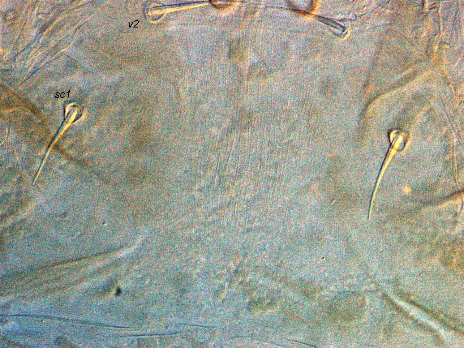
Fig. 10. Schizotetranychus baltazari adult male holotype - detail of the pattern of prodorsal striae.
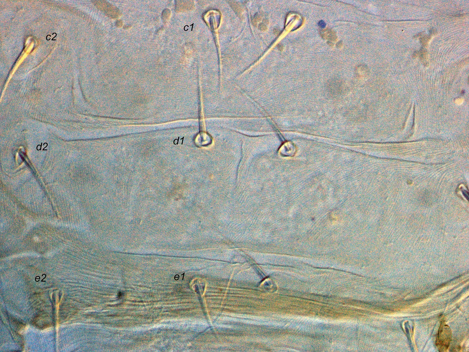
Fig. 11. Schizotetranychus baltazari adult male holotype - detail of dorsum, from setae c1-e1.
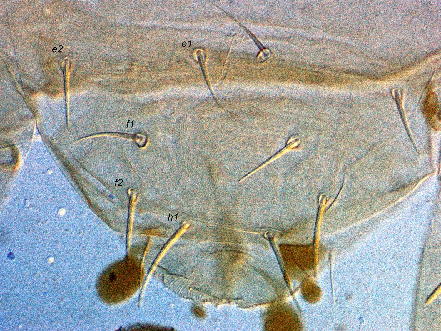
Fig. 12. Schizotetranychus baltazari adult male holotype - detail of dorsum, from setae e1-h1.

Fig. 13. Schizotetranychus baltazari adult male holotype - dorsal habitus.

Fig. 14. Schizotetranychus baltazari adult male holotype - detail of femur I.
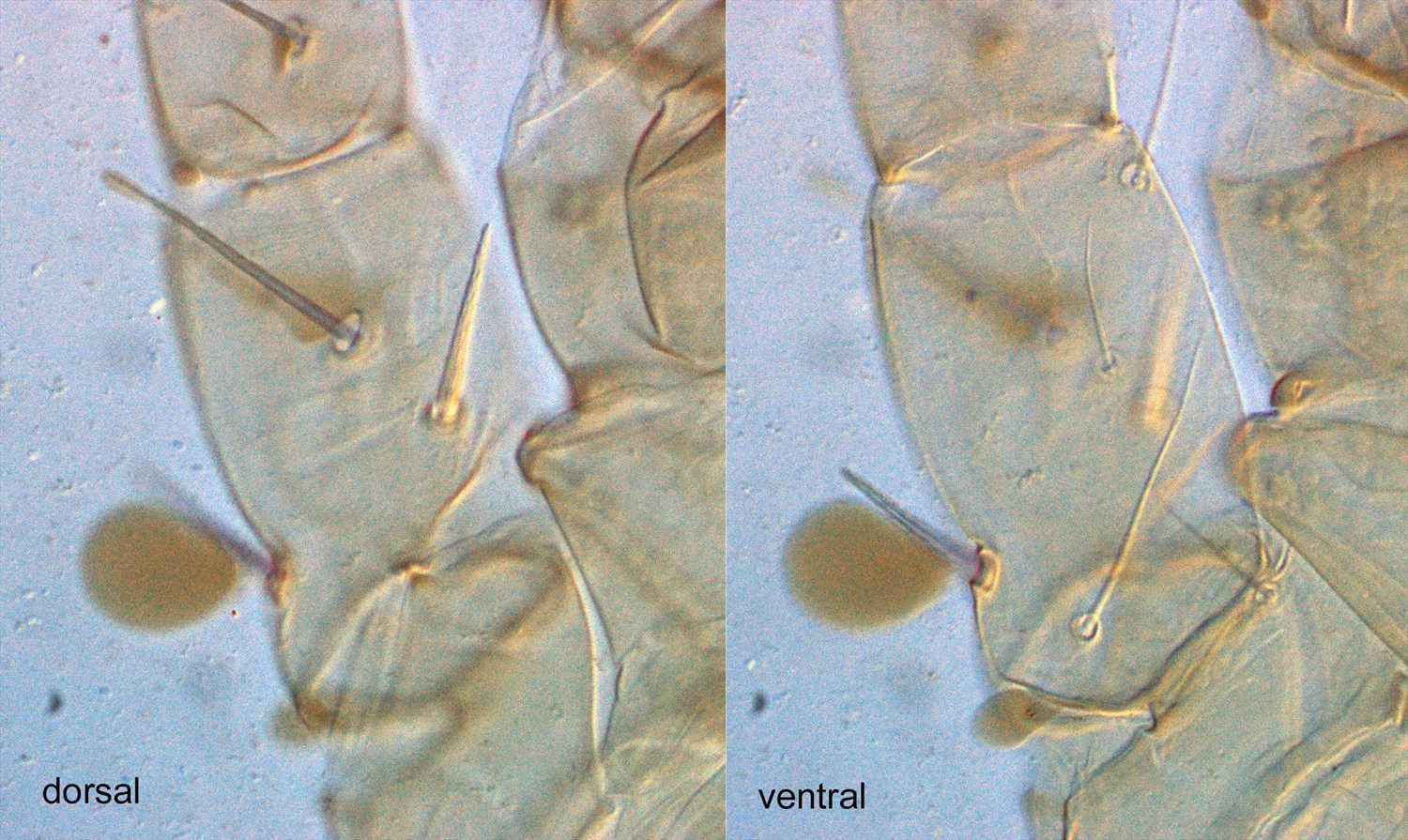
Fig. 15. Schizotetranychus baltazari adult male holotype - detail of femur II.
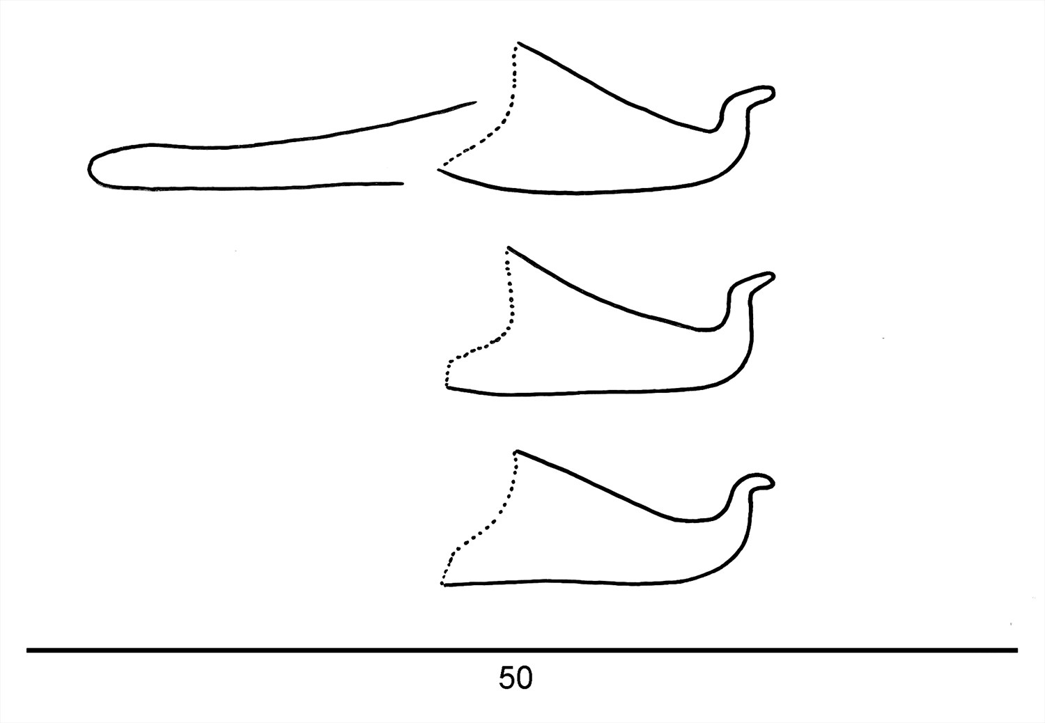
Fig. 16. Schizotetranychus baltazari adult male holotype - detail of aedeagus (at different focal points).
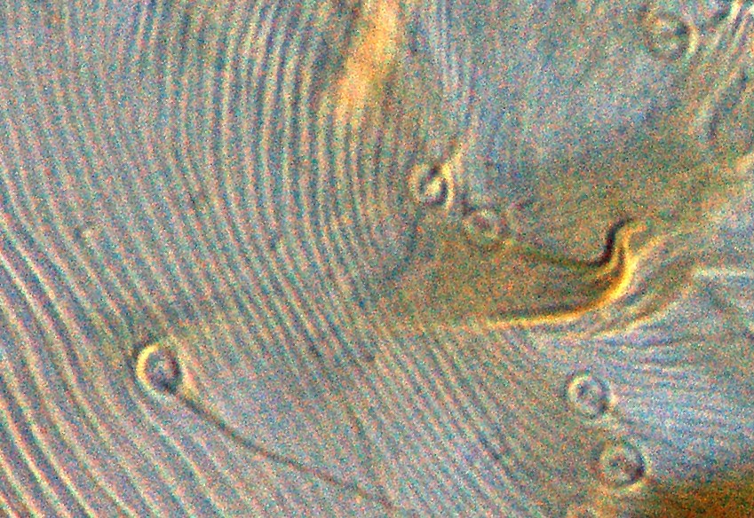
Fig. 17. Schizotetranychus baltazari adult male holotype - detail of aedeagus.
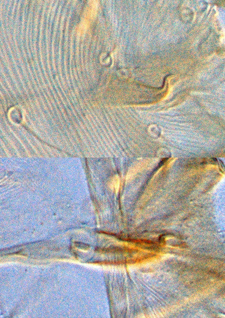
Fig. 18. Schizotetranychus baltazari adult male holotype (upper) and paratype (lower) - detail of aedeagus.
Material examined
types; non-types
Taxonomy
Subfamily Tetranychinae
Tribe Tetranychini
Common Name
none
Distribution
^^++NOT PRESENT IN AUSTRALIA
Burma, China, Hong Kong, India, *The Philippines, Taiwan, Thailand
Taxonomy Changes
Schizotetranychus baltazarae Rimando, Rimando (1962b)
Diagnosis
Female
- empodia I-IV = basal stalk (Fig. 8a) that is divided distally into two short, curved claws, thicker at base in lateral aspect; claws with a thin, curved dorsal hair, 3/4 length of claw (Figs 1, 2); minute proximoventral hairs present (Fig. 8a,b)
- pregenital striae fine, transverse, arching around genital flap; genital flap with mostly oblique striae (Fig. 3)
- tarsus I with the sockets of two tactile setae and one solenidion proximal to the socket of the proximal duplex seta
- tarsus II with the sockets of one tactile seta and one solenidion proximal to the socket of the duplex seta
- peritreme ending in short hook, like a golf club (Fig. 9)
- prodorsal striae longitudinal and distinctly broken, striae converging at a point just anterior to setae c1, forming a V-shape (Figs 4, 5)
- dorsal opisthosomal striae with strong lobes; longitudinal between c1-c1, d1-d1, e1-e1; transverse e1-h1 (Figs 6, 7)
- chaetotaxy for legs I-IV:
- femora 9, 6, 3, 3
- genua 5, 5, 3, 1
- tibiae 8(1+0), 5, 5, 5
- tarsi 15(3+3), 12(2+3), 8(1+0), 8(1+0)
Male
- empodia I-IV as in female (Fig. 8)
- peritreme same as female (Fig. 9)
- prodorsal striae fine, longitudinal, weak, widely spaced (Fig. 10)
- dorsal opisthosomal striae very fine, mixed patterns; with two weakly developed narrow transverse shields present, incorporating C-row and D-row (Figs 11, 12)
- coxae I enlarged, and leg I elongate (Fig. 13)
- femora I and II with enlarged thickened spur-like setae present (similar to that found on male Neonidulus) (Figs 14, 15)
- tarsus I with the sockets of three tactile setae and two solenidia proximal to socket of the proximal duplex seta
- tarsus II with the sockets of one tactile seta and one solenidion proximal to, and one tactile seta overlapping, the socket of the duplex seta
- chaetotaxy for legs I-IV:
- femora 9, 6, 3, 3
- genua 5, 5, 3, 1
- tibiae 11(4+0), 6(1+0), 5, 5
- tarsi 16(4+3), 12(2+3), 8(1+0), 8(1+0)
- aedeagus dorsally directed; no anterior or posterior projection (tip of projection curves posteriorly); dorsal margin of shaft at approx. 30° angle to ventral margin, abruptly bending dorsally at right angle forming dorsal projection; dorsal projection short, stout, finger-like, not tapering until very tip; dorsal projection curves posteriorly to form straight blunt tip; ventral margin of shaft straight (Figs 16-18)
Hosts
Citrus grandis, C. madurensis, C. medica, *Citrus nobilis, C. sinensis, Murraya koenigii (Rutaceae); Dioscorea sp. (Dioscoreaceae)
References
Migeon, A. and Dorkeld, F. (2006-2017) Spider Mites Web: a comprehensive database for the Tetranychidae. http://www.montpellier.inra.fr/CBGP/spmweb
*Rimando, L.C. (1962a) Four new species of spider mites of the genera Eotetranychus and Schizotetranychus (Tetranychidae, Acarina). The Philippine Agriculturist 14(10): 535-544
Rimando, L.C. (1962b) The tetranychoid mites of The Philippines. University of The Philippines, College of Agriculture Technical Bulletin 11: 1-52
Notes
Rimando (1962a) described and illustrated the aedeagus as much thinner and more strongly curved posteriorly to what is presented here of the holotype.
^^ ++ I consider Schizotetranychus baltazarae to be a suspected synonym of S. hindustanicus (Hirst, 1924). Until I examine and the holotype and paratypes of S. hindustanicus, the name S. baltazarae will be used here. However, this complicates the distribution records as specimens from northern Australia have been identified as S. hinustanicus.
Copyright © 2018. All rights reserved.