Click on images to enlarge
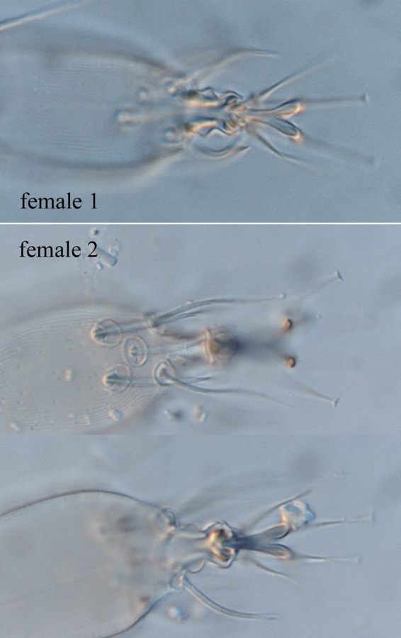
Fig. 1. Stigmaeopsis celarius adult female (non-type) - detail of divided empodia.
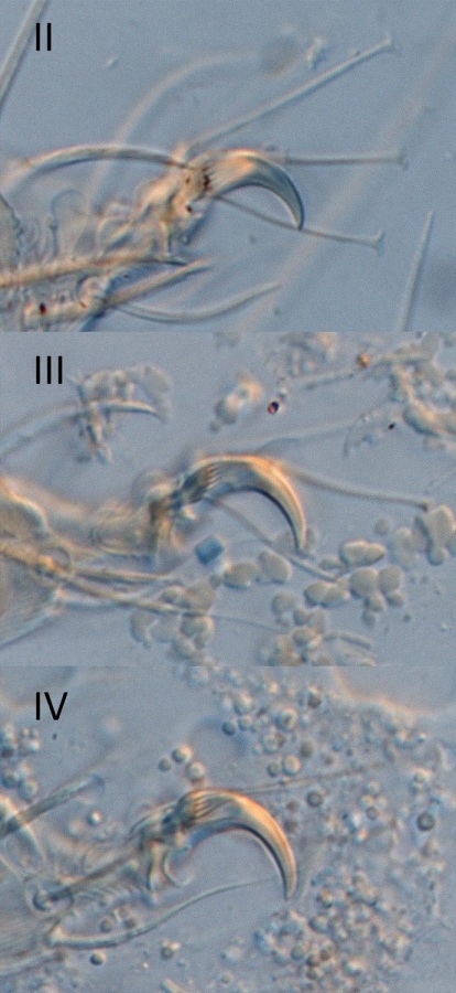
Fig. 2. Stigmaeopsis celarius adult female (non-type) - detail of empodia II-IV.
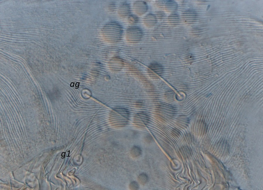
Fig. 3. Stigmaeopsis celarius adult female (non-type, USA) - detail of pregenital striae.

Fig. 4. Stigmaeopsis celarius adult female (non-type, Australia) - detail of pregenital striae.

Fig. 5. Stigmaeopsis celarius adult female (non-type, Australia) - detail of pregenital striae.
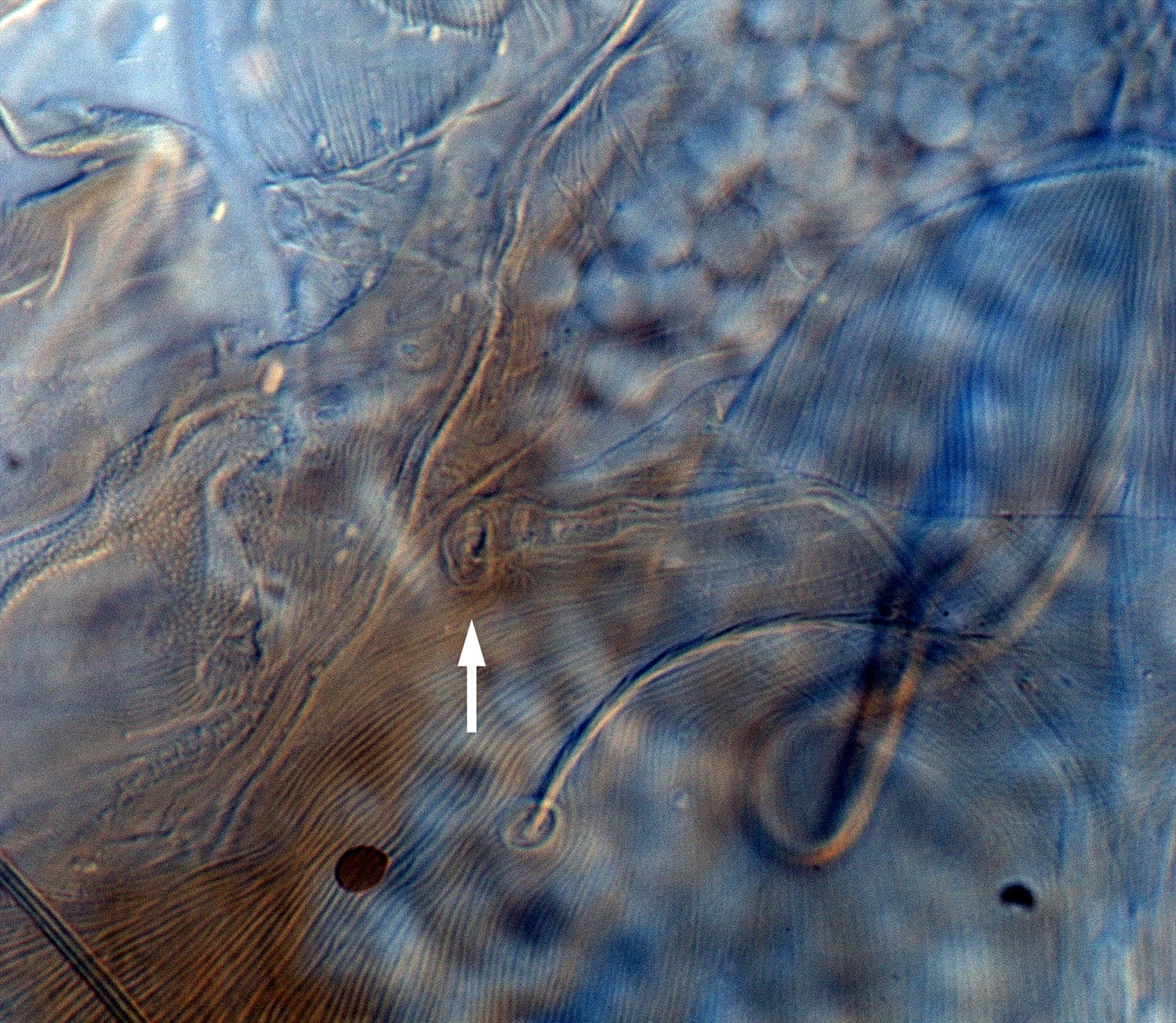
Fig. 6. Stigmaeopsis celarius adult female (non-type) - detail of peritreme (arrow indicates tip).

Fig. 7. Stigmaeopsis celarius adult female (non-type) - detail of peritreme.
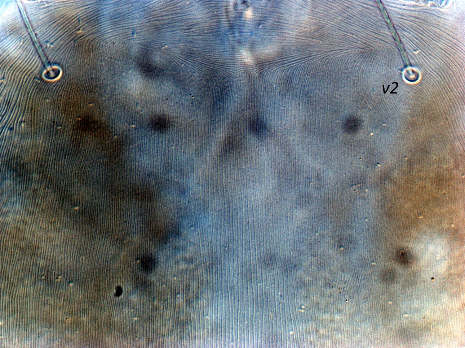
Fig. 8. Stigmaeopsis celarius adult female (non-type) - detail of pattern of striae on prodorsum.

Fig. 9. Stigmaeopsis sp. adult female - detail of palp tarsus, indicating 6 phaneres (3 eupathidia, one of which is the spinneret, 2 setae, one solenidion). e = eupathidia, set = seta, sol = solenidion). Redrawn from Saito et al. (2004).
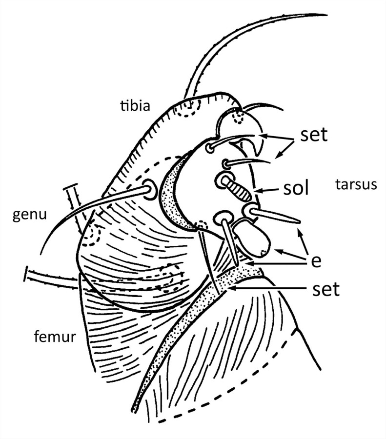
Fig. 10. Generalised tetranychid adult female - detail of palp, indicating 7 phaneres on the tarsus (3 eupathidia, one of which is the spinneret, 3 setae, one solenidion). e = eupathidia, set = seta, sol = solenidion). Redrawn from Lindquist (1985).

Fig. 11. Stigmaeopsis celarius adult female (non-type) - detail of dorsal opisthosoma.
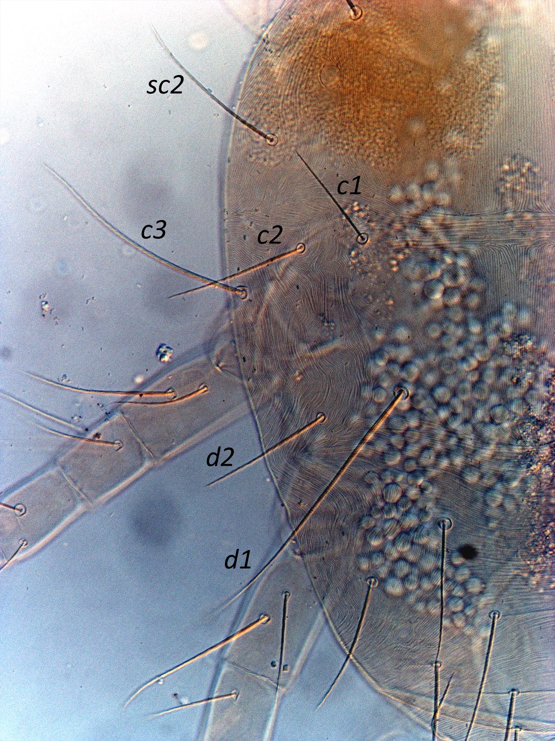
Fig. 12. Stigmaeopsis celarius adult female (non-type) - detail of dorsal opisthosoma (indicating long setae sc2, c3, d1).
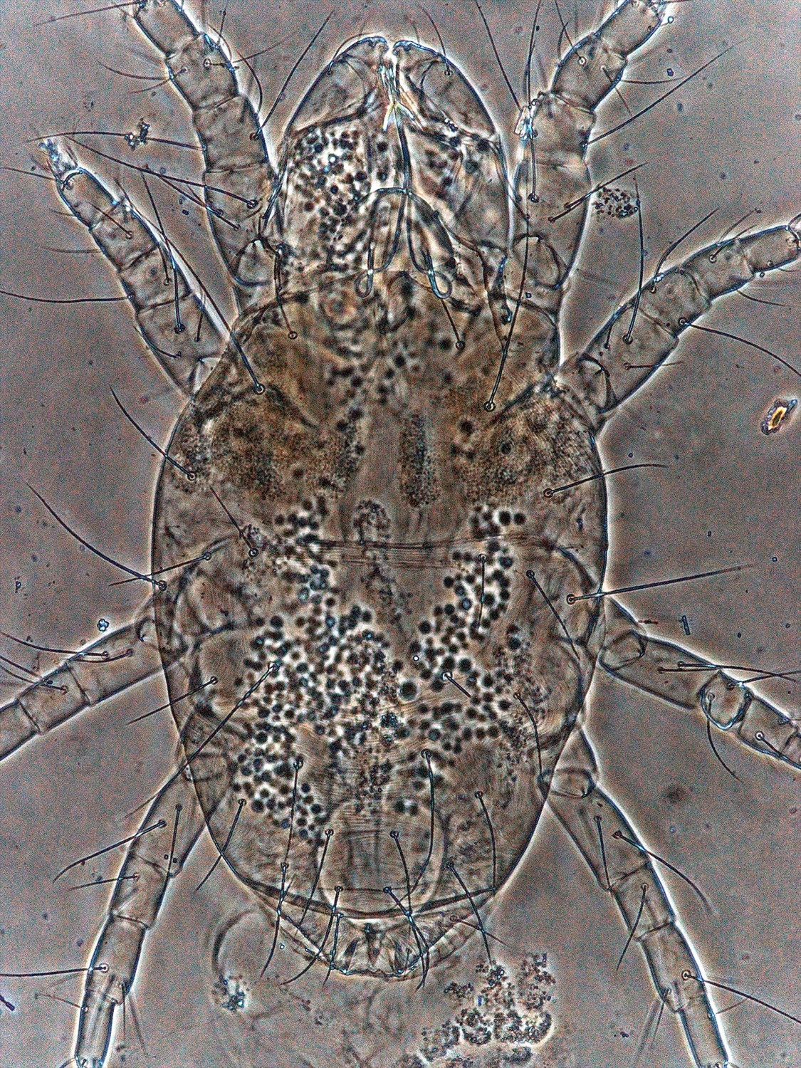
Fig. 13. Stigmaeopsis celarius adult female (non-type) - dorsal habitus.
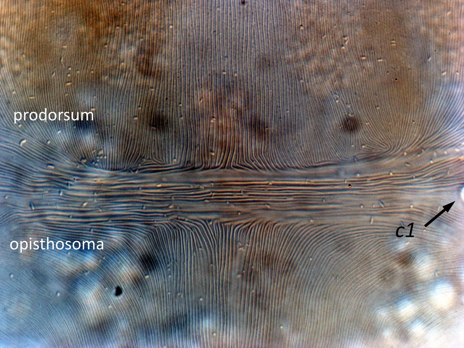
Fig. 14. Stigmaeopsis celarius adult female (non-type) - detail of pattern of striae at junction of prodorsum with opisthosoma (at level of setae c1; right side c1 partially visible).
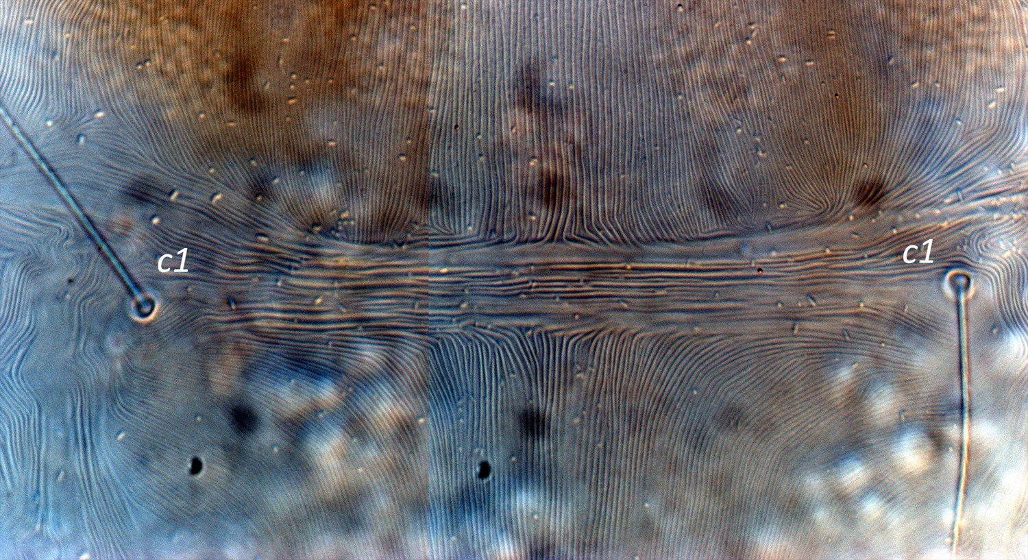
Fig. 15. Stigmaeopsis celarius adult female (non-type) - detail of pattern of striae at junction of prodorsum with opisthosoma (at level of setae c1).
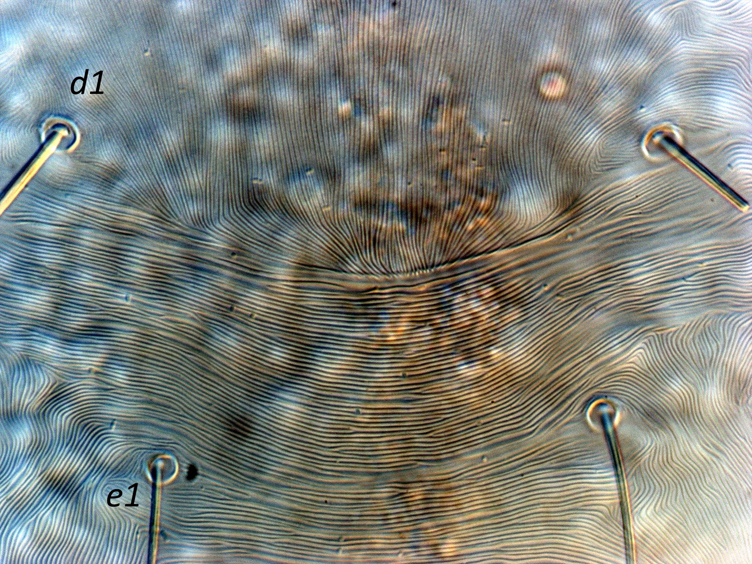
Fig. 16. Stigmaeopsis celarius adult female (non-type) - detail of pattern of striae on opisthosoma between setae d1 and e1.
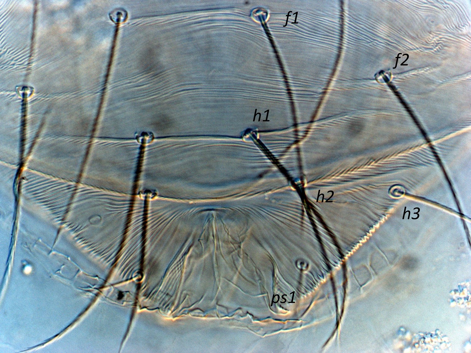
Fig. 17. Stigmaeopsis celarius adult female (non-type) - detail of setae on posterior dorsal opisthosoma.

Fig. 18. Stigmaeopsis celarius adult male (non-type) - detail of empodia I and IV.
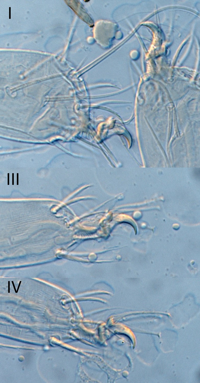
Fig. 19. Stigmaeopsis celarius adult male (non-type) - detail of empodia I, III and IV.
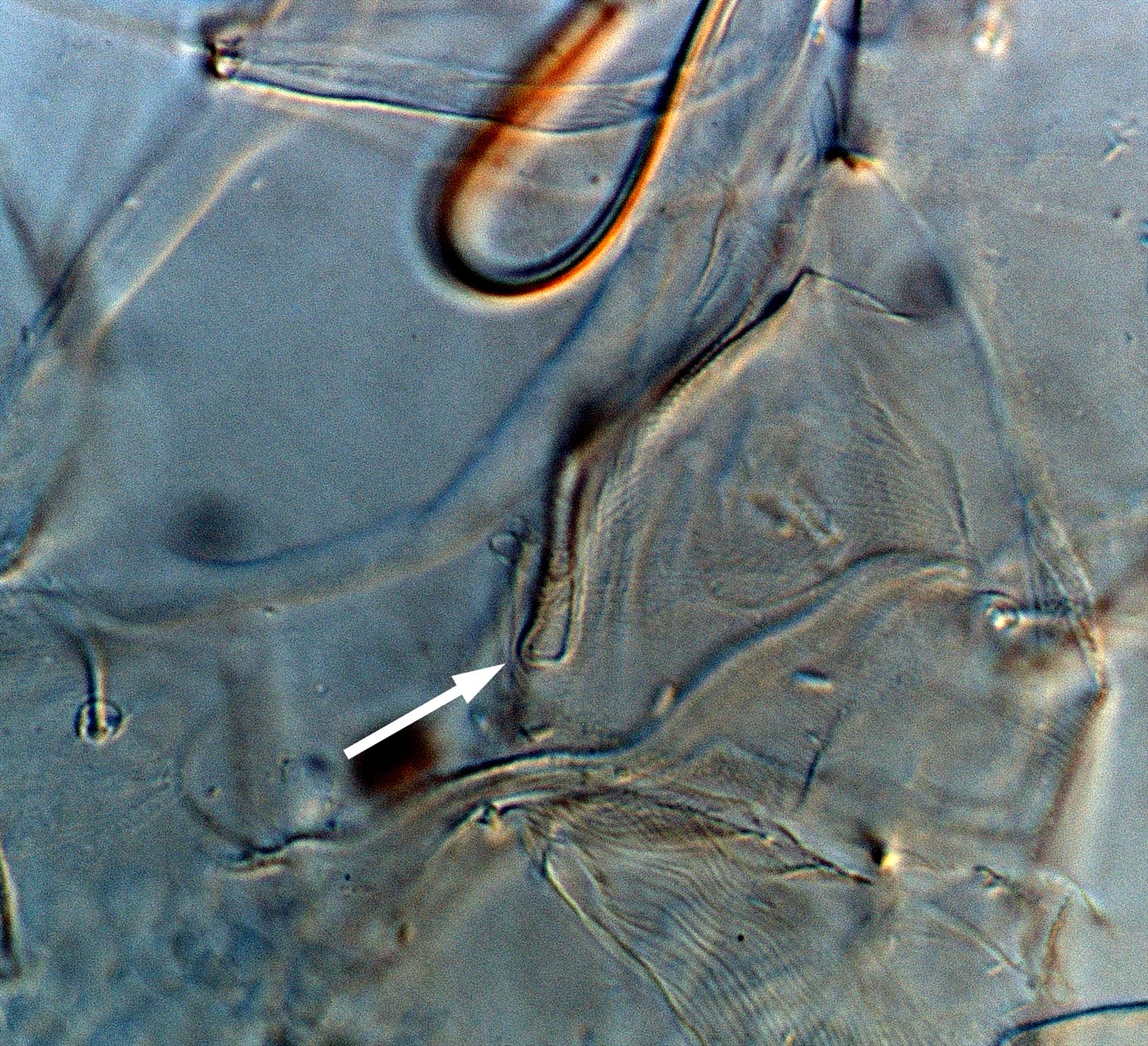
Fig. 20. Stigmaeopsis celarius adult male (non-type) - detail of peritreme (arrow indicates tip).
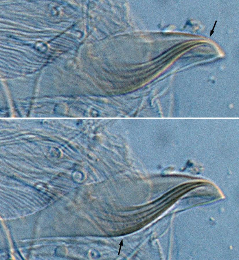
Fig. 21. Stigmaeopsis celarius adult male (non-type, France) - detail of aedeagus (not quite flat - arrows indicate tip in focus and shaft in focus).

Fig. 22. Stigmaeopsis celarius adult male (non-type, USA) - detail of aedeagus (not flat, arrow indicates tip).
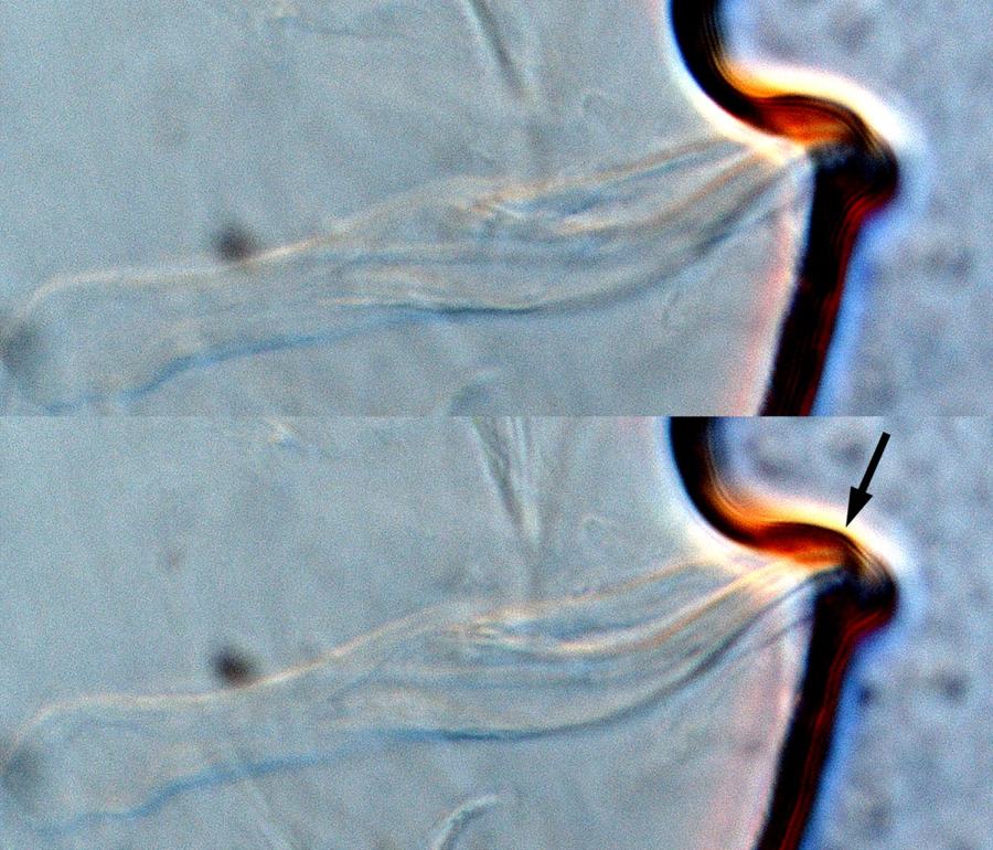
Fig. 23. Stigmaeopsis celarius adult male (non-type, France) - detail of aedeagus (not quite flat - arrow indicates tip).
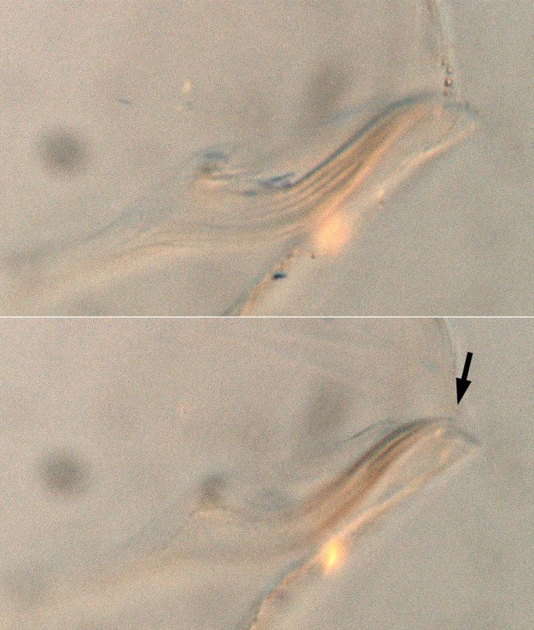
Fig. 24. Stigmaeopsis celarius adult male (non-type, France) - detail of aedeagus (not flat - arrow indicates tip).
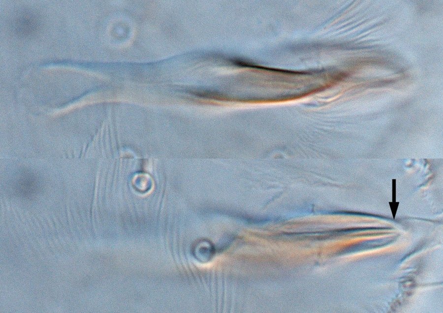
Fig. 25. Stigmaeopsis celarius adult male (non-type, Australia) - detail of aedeagus (not flat - arrow indicates tip).
Material examined
non-types
Taxonomy
Subfamily Tetranychinae
Tribe Tetranychini
Distribution
+Australia, China, France, Hawaii, Hong Kong, Japan, Korea, Okinawa Island, Taiwan, The Netherlands, *USA
Taxonomy Changes
Stigmaeopsis celarius Banks 1917
Schizotetranychus celarius (Banks) McGregor 1950
Schizotetranychus latitarsus Ewing 1917, synonymy McGregor 1950
Diagnosis
Female
- empodia I-IV divided into two "claws" (clearly visible from dorsal aspect, difficult to see in lateral aspect) (Figs 1, 2)
- pregenital striae transverse, but arching around anterior margin of flap (Figs 3-5)
- peritreme ending in simple unexpanded bulb (Figs 6, 7)
- prodorsum with longitudinal striae (Fig. 8)
- palp tarsus with only 6 phaneres (2 setae, 3 euphathidia, 1 solenidion) (Fig. 9) instead of 7 normally found on tetranychids (3 setae, 3 eupathidia, 1 solenidion) (Fig. 10)
- dorsal setae sc2, c3 and d1 much longer than other setae (Figs 11, 12)
- dorsal opisthosomal setae c1 inserted wide apart, close to c2 (Fig. 13)
- narrow band of transverse striae between setae c1-c1 (Figs 14, 15)
- dorsal striae longitudinal to weakly oblique between setae c1 & d1; striae transverse between setae d1 & h1 (Figs 16, 17)
- tarsus I with the sockets of one tactile seta and one solenidion proximal to, and one-two ventral tactile setae overlapping with, the socket of the proximal duplex seta OR with the sockets of one tactile seta proximal to, and one solenidion and one-two ventral tactile setae overlapping with, the socket of the proximal duplex seta
- tarsus II with the socket of one solenidion proximal to, and one-two ventral tactile setae overlapping with, the socket of the duplex seta
- chaetotaxy for legs I-IV:
- femora 8, 5, 3, 3
- genua 5, 4, 3, 2
- tibiae 8(1+0), 5, 5, 5
- tarsi 15(3+3), 12(2+3), 9(1+0), 9(1+0)
Male
- empodia I-IV divided into two "claws" (clearly visible from dorsal aspect, difficult to see in lateral aspect) (Figs 18, 19)
- peritreme ending in simple unexpanded bulb (Fig. 20)
- dorsal setal lengths similar to female
- tarsus I with the sockets of one tactile seta and one solenidion proximal to, and one solenidion and two ventral tactile setae overlapping with, the socket of the proximal duplex seta
- tarsus II with the socket of one solenidion proximal to, and two ventral tactile setae overlapping with, the socket of the duplex seta
- tarsi I-IV with 16(4+3), 12(2+3), 9(1+0), 9(1+0)
- aedeagus dorsally directed, with anterior and posterior projections absent; dorsal and ventral margin of shaft m ore or less straight and parallel; dorsal projection at 40-45° angle to main axis of shaft, tapering to a finely truncate tip (Figs 21-25)
Hosts
Approximately 20 recorded species of host plant, including mainly Poaceae: Bambusa sp. (Poaceae), Ficus stipulata (Moraceae), Oryza sativa, Phyllostachus makinoi, P. nigra, P. reticulata, Pleioblastus shibuyanus, *Pseudosasa japonica, Saccharum officinarum, S. spontaneum, Sasa kurilensis, Sas. nipponica, Sas. senanensis (Poaceae)
Biology
This species is a common pest of bamboo in Japan and an occasional pest of rice and sugarcane. Like its relatives in Schizotetranychus, these mites live in discrete colonies under dense webbing, located on the underside of leaves. Their feeding causes chlorotic spots that can be seen on the upper surface of the leaves as white spots.
References
Baker, E.W. and Tuttle, D.M. (1994) A guide to the Spider Mites (Tetranychidae) of The United States . 347 pp. Indira Publishing House, Michigan.
*Banks, N. (1917) New mites, mostly economic (Arach. Acar.) Entomol. News 28: 193-199
Ewing, H.E. (1917) New species of economic mites. J. Econ. Entomol. 10: 497-501
+Gutierrez, J., and Schicha, E. (1983) The spider mite family Tetranychidae in New South Wales. International Journal of Acarology 9(3): 99-116
Jeppson, L.R., Keifer, H.H. and Baker, E.W. (1975) Mites injurious to economic plants. 614pp. Berkeley, University of California Press.
McGregor, E.A. (1950) Mites of the family Tetranychidae. American Midland Naturalist 44: 257-420
Migeon, A. and Dorkeld, F. (2006-2017) Spider Mites Web: a comprehensive database for the Tetranychidae. http://www.montpellier.inra.fr/CBGP/spmweb
Notes
Copyright © 2018. All rights reserved.