


All true fungi belong to the Kingdom Eumycota (Greek, eu = true; mykes = a fungus). The fungal kingdom includes rusts, smuts, moulds and mildews, as well as the 'higher fungi'. The two divisions or groups of higher fungi are Basidiomycota (basidiomycetes) and Ascomycota (ascomycetes) and each division is defined by the way in which the fungus produces its spores. For mycologists, the word 'division' has the same meaning as the word 'phylum' has for zoologists. Both mean a very large grouping of organisms which are similar in some very basic characteristics.
Ascomycetes
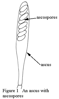
If a fungus belongs to division Ascomycota, then at some stage during its lifecycle it must produce a specialised cell called an ascus (plural: asci). Asci have several shapes, but the most common is like an elongated club. Usually eight spores are formed inside the ascus, but this number can vary from one to several hundred, depending on the species (See Figure 1 for a diagram of an ascus with ascospores).
When the spores are ripe the ascus may release them in several ways. Some asci develop a line of weakness (almost like a zip fastener) around the top of the ascus which then allows a small cap to break away and the spores are then squirted out. Another method is for a small pore to develop at the top of the ascus through which the spores are released. A third process involves the asci breaking apart.
Most people are not familiar with many ascomycetes but one common species is known in Victoria and Tasmania as the 'beech orange' (Cyttaria gunnii) which forms orange, golf ball-shaped fruiting bodies on branches of Southern beech or myrtle (Nothofagus cunninghamii). Another ascomycete used in gourmet cooking is the morel (Morchella spp.) which is highly prized as an edible fungus in Europe and America. Morels occur infrequently from Tasmania to southern Queensland. A third group of very highly prized ascomycetes are the truffles, e.g. Tuber melanosporum (Black Truffle) and T. magnatum (White Truffle). Truffles are not native to Australia.
Ascomycetes in this key include the genera listed below. The scientific names of the families concerned can be found in any suitable reference work.
Disc or cup fungi of several families: Aleuria, Chlorociboria, Cyttaria and Scutellinia.
Earth tongue fungi: Leotia.
Stroma-developing fungi of several families: Cordyceps, Daldinia and Xylaria
.
Basidiomycetes
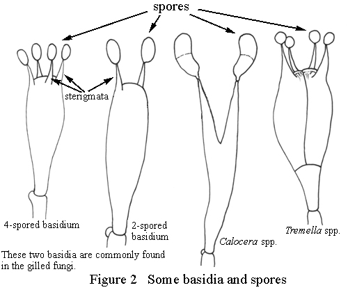
If a fungus belongs to division Basidiomycota, then at some stage during its lifecycle it must produce a specialised cell called a basidium (plural: basidia). Basidia have a variety of forms, but usually they resemble an elongated club with short spikes (sterigmata) growing from the top. One spore is produced at the tip of each sterigma. Some basidia produce only two spores, others produce up to perhaps eight; however, the most common number of spores is four (See Figure 2 for a diagram of various types of basidia).
Most people are familiar with some members of this division: the mushrooms and toadstools. Other members include the puffballs, earth stars, jelly fungi, coral fungi and all bracket fungi. There are also a number of microscopic fungi that belong to this division such as the rusts and smuts of cereal crops. Most of the commercial edible mushroom crops that are now finding their way into the supermarkets are basidiomycetes. Examples are new-comers such as the oyster mushroom (Pleurotus ostreatus) and the shiitake or Japanese mushroom (Lentinula edodes).
Basidiomycetes in this key include the genera listed below, and the scientific names of the families concerned can be found in any suitable reference work.The genus Schizophyllum is placed in the gilled fungi for convenience as it produces gills. Recent research, however, indicates that it may be closer in its relationships to the pored fungi.
Jelly fungi: Auricularia, Pseudohydnum and Tremella.
Stinkhorn fungi: Asero, Colus, Dictyophora, Lysurus, Mutinus, Phallus and Pseudocolus.
Puffball fungi, earth stars and close relatives: Calostoma, Geastrum, Lycoperdon and Pisolithus.
Bracket fungi: Ganoderma, Laetiporus, Phellinus, Piptoporus, Pycnoporus, Trametes and Tyromyces.
Leathery or stereoid fungi: Cymatoderma and Stereum.
Toothed fungi: Hericium and Hydnum.
Coralloid fungi: Clavaria, Clavulinopsis and Ramaria.
Boletoid or pored fungi related to gilled fungi: Boletellus.
Cantharelloid fungi very close to gilled fungi: Cantharellus.
Gilled fungi or agarics: Agaricus, Amanita, Anthracophyllum, Armillaria, Calyptella, Conchomyces, Coprinus, Crepidotus, Cyptotrama, Descolea, Entoloma, Filoboletus, Hygrocybe, Lactarius, Lentinellus, Lentinula, Leucocoprinus, Macrolepiota, Marasmius, Mycena, Omphalotus, Pholiota, Pluteus, Russula, Schizophyllum, Stropharia, Xeromphalina and Xerula.

Tests for Edible Fungi from Folklore
The Fungal Toxins
Introduction
Many species of fungi are used regularly as food by humans. Each Autumn, thousands of people go into the fields and woodlands of Europe in search of edible fungi which are collected and then either cooked immediately, or preserved in some way for later use. Among the most prized species gathered are the chanterelle, the giant puffball, ceps and the shaggy inkcap, all of which would not be recognised by most Australians as edible despite the fact that some of these species occur in Australia.
Some Asian higher fungi are now regularly seen fresh, canned or dried in supermarkets or Asian food stores and include such species as the shiitake, the enoki fungus, straw mushrooms and Jews ears. The oyster mushroom is a European fungus that now appears very frequently in a fresh state.
In Europe, there is an ethnic tradition in almost every country which ensures that wild fungi will be collected for food. This tradition probably dates back to times when, during severe winters, every item of stored food contributed to survival. The crop of fungi which appeared every Autumn was a food source that could not be ignored, and so local knowledge of the edible fungi was carefully learnt and passed on from parents to children. This store of knowledge was originally learnt in a rather brutal way: if the fungus was not toxic and was good to eat, one survived, otherwise one became ill or even died.
Despite the local knowledge of the edible fungi in Europe, things still do go horribly wrong and at least 100 people die each year from eating toxic fungi. The most commonly eaten poisonous species is the Death Cap, Amanita phalloides. This fungus produces poisons which attack liver and intestinal function and, without swift hospital treatment, the fatality rate is more than 90 per cent. The Death Cap does not occur naturally in Australia; unfortunately, however, it was accidentally introduced into Canberra and Melbourne as a mycorrhizal species on the roots of oak trees and is now well established. There has already been one Australian fatality definitely attributed to this species.
Despite the proliferation of new fungal foods, Australians in general are very cautious when considering wild fungi and rightly so. Australians do not have a tradition of eating wild mushrooms and consequently most of our fungi have not yet been tested for their edible qualities. Problems also arise when migrants or visitors from Europe walk through Australian forests and believe that they recognise fungal species that are edible. Unfortunately, this generally is not the case. There are large numbers of Australian species which superficially resemble European species, but are in fact quite different. This similarity has lead to some past cases of fungal poisoning.
Anyone intending to eat wild fungi MUST have the fungi identified by a qualified mycologist before considering using them for food. Experimentation with unknown fungal species is extremely dangerous and can even be fatal. In addition, many people are allergic to even well known edible species so that a great deal of caution must always go hand in hand with trying a new species.
Tests for Edible Fungi from Folklore
There is no known, infallible test for determining if a fungus is edible or not. If anyone intends to collect wild fungi for food, then the characteristics of the edible species must be learnt and recognised. There are some traditional ways of testing a fungus for poisons and the following list is included with examples of where each one fails.
|
'A fungus is edible if the skin of the cap peels'. |
This is true for the field mushroom, but is also true for the Death Cap |
|
'If the taste is pleasant, it is safe to eat'. |
Chlorophyllum molybdites is very poisonous in the raw state, and its victims have stated that it is delicious to eat when uncooked. |
| 'If a silver coin is placed in the cooking fungus, the silver will draw the poison out and neutralise it or the silver will blacken if the mushrooms are poisonous'. | Silver has no ability to deal with any fungal poison. The only way it will most certainly blacken is if eggs are part of the dish. This test is pure medieval alchemy. |
| 'If there is evidence of animals having eaten the fungus then it will be suitable as food'. |
Unfortunately, slugs enjoy a meal of the Death Cap and rabbits can eat small amounts with impunity. |
| 'If the fungus changes colour when cut, it is toxic'. |
Some toxic fungi such as the Yellow Staining Mushroom do change colour when cut, but so do many very good edible species among the boletoid fungi. |
| 'Brightly coloured fungi are poisonous.' | This is quite untrue; the Chanterelle, one of Europe's most prized edible fungi, is brilliant egg-yolk yellow, while Blewitts are bright lilac and are an excellent edible species after cooking. |
| 'Blanching in salty/vinegared water makes even the most toxic species edible'. | This extremely dangerous fallacy was apparently suggested by the French naturalist Fabre. The Death Cap is completely unaffected by blanching. |
| 'Drying removes the toxins' | This is true for one European species, but is not true for the Death Cap or those species which contain the kidney destroying toxin orellanine. |
| 'Fungi growing on rubbish tips are certain to be poisonous'. | This test was first suggested by the Roman, Pliny the Elder (2379AD). The deliciously edible shaggy caps grow under precisely these conditions. |
| 'Poisonous mushrooms cause egg-white or milk to curdle'. | Some may do so, but then so do two of the most highly prized European edible species, Caesars Mushroom (Amanita caesarea) and the Cep (Boletus edulis). |
The Fungal Toxins
The fungal toxins fall into several groups. Only brief details will be given here.
| Amatoxins | This group of poisons was first recognised from Amanita phalloides, but amatoxins also occur in several other related species. Amanitin attacks the liver and its functions. It is exceptionally dangerous as the liver tries to remove the toxin by ejecting it into the bile. Unfortunately, the bile goes into the intestine from where the toxin is reabsorbed. Hospital treatment is essential. Amatoxins have a delayed effect and symptoms typically do not begin for four to eight hours after ingestion. There is violent vomiting and stomach pain and, if untreated, death occurs in about four days after the patient lapses into a coma. |
| Muscarine | This toxin is dangerous but rarely fatal. It was first recognised from the Fly Agaric (Amanita muscaria), but many other fungal species have the toxin in much greater amounts. The symptoms are very distressing and include watering eyes, runny nose, the production of large amounts of saliva, heavy perspiration, blurred vision and constricted pupils. There may be some stomach pain. Hospital treatment is essential and there is an excellent antidote (atropine). |
| Orellanine | This very dangerous toxin has only been fully understood in the last 1020 years. It occurs in some members of the genus Cortinarius and, in particular, those species that are small and brown. The poison attacks and destroys the kidney cells. If enough has been eaten, the patient completely loses kidney function and survival requires either dialysis or a kidney transplant. The main symptoms of the poison may not appear for as much as two weeks after ingestion, by which time the meal with the toxic fungi has been forgotten. |
| Coprinus-Alchohol Toxicity | This curious effect occurs only if Coprinus atramentarius or one or two other very closely related species are eaten during a meal where alcohol is served. Without alcohol, there are no symptoms. The alcohol allows a poison in the fungus to be absorbed, and the possible effects include nausea, vomiting, rapid pulse, dizziness and violent flushes of the skin. |
| Psilocin and Psilocybin | These toxins produce hallucinations. The poisons interfere with the central nervous system and produce fantasy, slurred speech, uncontrollable laughter, dizziness, distorted vision, etc. The effects wear off after about 68 hours depending upon the amount eaten. Flashback phenomena, where the patient can suddenly start hallucinating, even though no fungus was recently ingested, do occur. |
| Gastrointestinal Toxins | A variety of species produce toxins which cause symptoms of gastrointestinal irritation including vomiting, nausea, pain and diarrhoea. Depending upon the species, the symptoms may be limited to vomiting and ejection of the offending fungus or very severe pain with associated diarrhoea and dehydration to the extent that hospitalisation and fluid replacement are essential for survival. |

Introduction
In some ways, a fungus behaves like a green plant: it stays in the same place and it produces spores which have the same function as seeds in higher plants. About 100 years ago, botanists placed the fungi in the plant kingdom because of these similarities, but today many consider that the fungi should be placed in at least three separate kingdoms because they are so very different from true plants. These differences are often microscopic and biochemical and will not be dealt with here.
The visible toadstool or mushroom is not the whole fungus, it is only the fruiting body, and it has a similar relationship with the fungus mycelium as an apple has with its parent tree. The bulk of the fungus is hidden inside the material (substrate) from which the fruiting body has appeared.
The fungus body is called a mycelium and it resembles a mass of very soft, fine threads. Many mycelia are white but they may also be red, orange, green, brown and black. Each individual thread is called a hypha and the hyphae carry out all the activities needed for fungal growth. Fungi grow well on human food materials and mouldy bread or fruit may be covered by a mass of grey-white mycelium and a number of fruiting bodies consisting of vertical threads, each capped with a tiny black or brownish ball containing spores. The fruiting bodies resemble pins and mycologists sometimes refer to this group of fungi as 'pin-moulds'. The green moulds that may appear on oranges are frequently species of the genus Penicillium, the group of moulds that produces penicillin.
The Higher and Lower Fungi
The fungi in this key are often called higher fungi or macrofungi. To mycologists the first term means that these fungi are considered to be more advanced and complex in their development than some other fungal groups while the second term means that they are easily seen with the naked eye. Most people rarely notice the lower fungi or microfungi although they are frequently affected by them and often use the microfungi (or their products) in daily life.
One of the microfungi, Phytophthora infestans, causes the disease potato blight and was responsible for the deaths of more than 500,000 Irish people during the late 1840s when it wiped out the potato crops of Ireland and caused mass starvation. Many of the microfungi cause plant diseases such as wheat rust or rose black spot, but they are also helpful in plant litter decay and are responsible for some extremely important economic processes. Many types of cheese need moulds to assist their development, our antibiotics mostly come from microfungi, organ transplants are impossible without a substance (cyclosporin) produced by one of the microfungi and yet another, a yeast, is an important ingredient in bread-making. Yeast is also essential for the production of wines, beers and spirits.
The higher fungi include all those bushland fungi that are easily seen with the naked eye and which are normally called 'mushrooms' and 'toadstools'. These fungi produce quite complicated fruiting structures and also show some microscopic characteristics which lead mycologists to believe that they are far more advanced in their life styles than the more primitive lower fungi.
The higher fungi are critically important in plant litter recycling but also have a significant role in the health of the forests by forming partnerships with the trees (for a detailed description see Mycorrhizal Relationships). The higher fungi are also very important commercially as food crops. Many species, including the ordinary field mushroom, are eaten, and some can now be found on Australian supermarket shelves. Two that are often sold fresh are the Japanese shiitake, Lentinula edodes, and the European oyster mushroom, Pleurotus ostreatus. The paddi or rice straw mushroom, Volvariella volvacea, is commonly found canned in the Asian food section.
Another important aspect of the higher fungi is their role in animal food resources. Not enough is known about the part many of these fungi play, but higher fungi are consumed by small mammals, reptiles insects and other invertebrates. Often the animal acts as a spore dispersal agent for the fungus.
Growth and Reproduction
A mycelium increases its size by growth at the tips of the hyphae. The hyphae absorb food and process it, and when conditions are right, they form the reproductive structure. In the forest, the growing tip of a fungal hypha encounters materials that are suitable as food sources, e.g. leaves, branches or logs. Dead plant material is quite hard and bacteria cannot break it apart easily, but fungi have two advantages: they produce specialised chemicals (enzymes) and their hyphae can easily penetrate enzyme-softened tissues. The fungal enzymes break apart large molecules such as cellulose with which the plant makes its cell walls. The chemical changes produced by the enzymes cause the wood to soften and the hypha then grows into the softened wood. Other enzymes are now produced which break down the cellulose into simple sugars which are absorbed by the hyphae. So, little by little, the mycelium steadily invades the dead wood and uses it for food. Eventually, when all the nourishment is exhausted, all that will be left will be a completely rotted log that is being attacked by borers and other insects and within a few years, the whole log will have returned to the compost of the forest floor. There is no doubt that bacteria also act as decomposers for plant material; however, the fungi appear to be the primary colonisers for hard organic materials such as fallen logs.
For most fungi, precise combinations of temperature, water and food availability are required to induce the production of fruiting structures. Many seem to require a small drop in temperature along with sufficient moisture and so there is always a flush of mushrooms and toadstools in autumn. The fungus has been storing food for a considerable time during summer and when conditions are favourable, it reacts very quickly to put many hyphae together and produce the toadstool that seems to appear almost overnight. In fact, the fungus has probably been waiting with an immature and unexpanded fruiting body for several weeks, but the sudden appearance of the fruiting body is often startling to many people.
Once the fruiting body is developed, it begins to produce spores. Spores are tiny cells capable of producing a new fungus, but they are not constructed in the same way as the seeds of higher plants. The spores of commercial mushrooms are so small that about 100150 of them placed end-to-end would be one millimetre. A mushroom can produce millions of them every hour and one estimate suggests that over a three day period, an ordinary field mushroom produces over 700 million spores. This pales to insignificance when compared a giant puffball, estimated to produce 5000 million spores.
So, why are we not overwhelmed by fungi? The answer is that spores usually do not survive very long as they can carry only a minute amount of food, and they die quickly unless they find a suitable substrate. Of the many millions of spores that are produced in autumn, only a very few will survive and grow to make a new fungus.
Fairy Rings
For hundreds of years, people have wondered at the sight of fairy rings. These are either bare circles of ground or rings of mushrooms, toadstools or puffballs that appear in grassy paddocks. Many explanations have been suggested but the answer is very simple. When a spore germinates, it needs food. Assuming it is a grassland species, it needs dead grass or humus. The mycelium grows radially in search of suitable substrates and so forms a circular colony. Sooner or later, the food runs out at the centre and the fungus dies, but the edges of the circle are still active and so a ring shape forms. When conditions are right, fruiting structures are produced where the fungus is actively growing, i.e. at the edges of the ring. An image of a fairy ring is attached. The shape of the ring is very clearly defined and the fertiliser effect of the fungal wastes shows in the very rich growth of the grass at the centre of the ring.
Luminous Fungi
Some fungi glow in the dark and this occurs due to the combination of two chemicals which produce light when they make contact. The chemicals are simple to make and can be bought in speciality shops where they are called 'glow sticks'. They contain two liquids which when mixed cause the stick to glow with a greenish light for several hours.
The purpose of luminescence in fungi is uncertain, but it may be important for their dispersal. One of our common glowing fungi, Omphalotus nidiformis, produces a greenish white light, has a strong odour and is highly attractive to the large land snails in rainforests. It is quite possible that it is the combination of light and smell which attracts the snails. After feeding on the fungus, the snails carry the spores in their intestines and possibly disperse them in their droppings. There is a photograph of a tree covered in Omphalotus nidiformis showing it by day and by night in the taxon information section.
Another species of luminous fungus, Mycena chlorophos, is a small, greyish and viscid toadstool with a conical cap that is often found on the dead fronds at the base of palm trees. It has a brilliant green light and is much brighter than Omphalotus nidiformis. It appears in quantity during the warm, wet summer months northwards from Sydney. During its growing season, there are many telephone calls to museums and herbaria concerning discoveries of glowing toadstools.
Vegetable Caterpillars
These curious fungi have long been known to scientists and also to the Chinese who use certain species for medicinal purposes. They result from the attack of a parasitic fungus which invades the bodies of very large caterpillars when they burrow underground to pupate. The fungi belong to the genus Cordyceps (an ascomycete), but not all species of Cordyceps attack these large caterpillars, and others may be found on the pupae of small moths or butterflies, and the bodies of wasps or ants. Australia is particularly fortunate in that it has the largest species in the world, Cordyceps taylori, which produces above-ground fruiting structures that resemble the antlers of a deer and may be up to 20 cm high.
The life cycles of most of these species have yet to be understood. However, for those species that attack the large burrowing caterpillars, it is known that the fungus attack occurs once the caterpillar has completed at least part of its burrow. It is not known whether the caterpillar has already eaten the fungus spore which remains dormant until triggered by the hormonal changes in the caterpillar as it undergoes metamorphosis, or whether the spore is in the soil and makes contact with the caterpillar while it is burrowing. It is certain, however, that the fungal attack is very rapid and the caterpillar is killed extremely quickly. The fungus then expands throughout the body of the caterpillar and consumes all its tissues. Eventually, all that remains are the outer cuticle and some of the harder chitinous parts such as the jaws and feet. The interior of the caterpillar is a dense mass of fungal hyphae. The fungus waits until climatic conditions favour spore dispersal and the fruiting body then grows up through the soil. Cordyceps gunnii produces green clubs about 10 cm high, while in C. hawkesii they are brown and about the same size. A common but often overlooked species, C. militaris, produces bright orange clubs about 23 cm high.
Figures
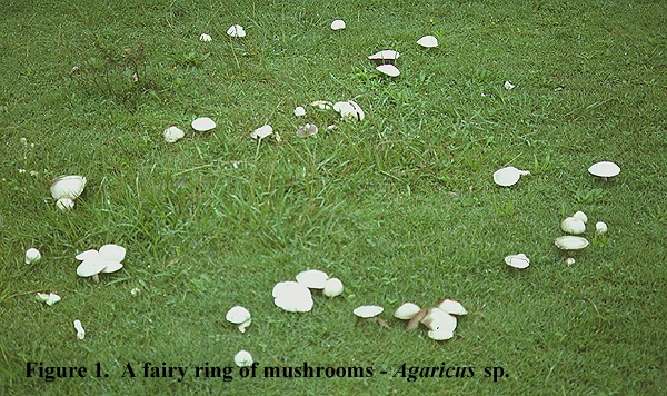
1. A fairy ring consisting of fruiting bodies of a species of Agaricus (mushroom). The ring is a little uneven but shows the more or less circular pattern being developed.
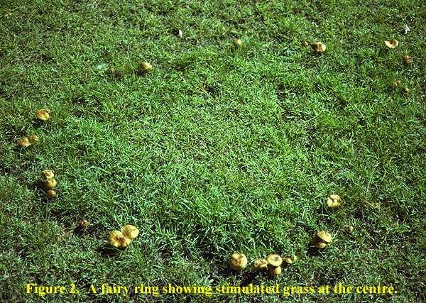
2. A fairy ring of an unknown species of Marasmius. This ring shows the growth stimulant effect of the fungus products; the grass inside the ring is more lush than outside.
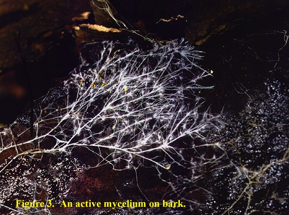
3. Part of a fungal mycelium growing on the undersurface of rotting bark. The mycelium is radiating outwards by successive branchings and also has thickened hyphal strands (rhizomorphs) which carry nutrients through the fungus.
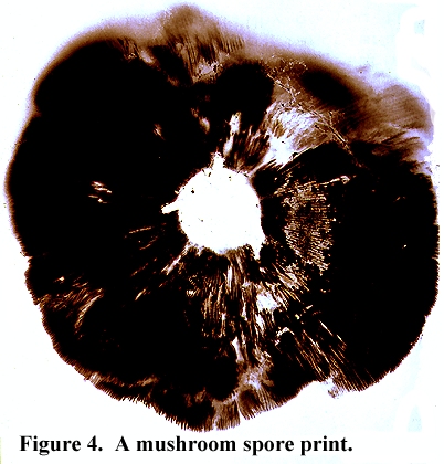
4. A spore print produced from a mushroom (Agaricus sp.). The spore print contains about 200 million spores and was produced by leaving a mushroom cap placed gills downwards on a piece of white paper overnight.
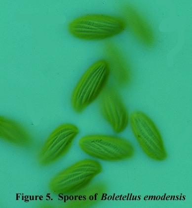
5. Spores of Boletellus emodensis. Each spore is about 18 m long. The tiny cross striations between the long axis striations are clearly visible.
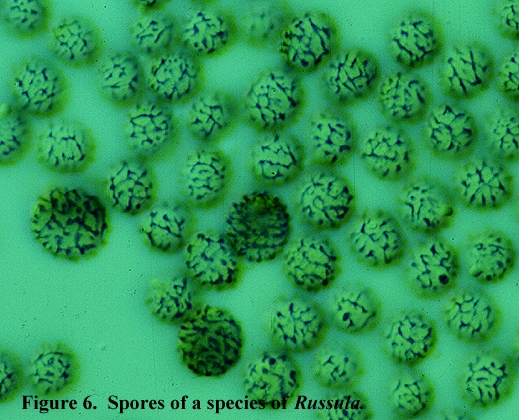
6. Spores of a species of Russula. Each spore is about 10 m in diameter. A stain has been used to show the ridges and warts on the surface of the nearly globose spores.
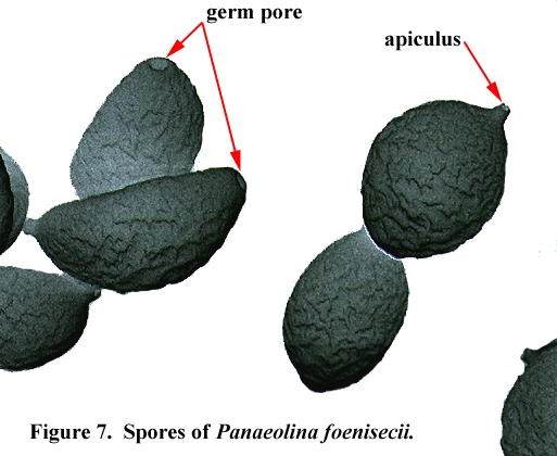
7. Spores of Panaeolus foenisecii. These spores were photographed under a scanning electron microscope and they about 12.5 m long. The photograph shows the wrinkled surface of the spores and a germ pore at one end. The germ pore is a weak point in the cell wall where the germinating fungus will emerge from the spore. At the other end of the spore is the apiculus, the point of attachment to the parent basidium.
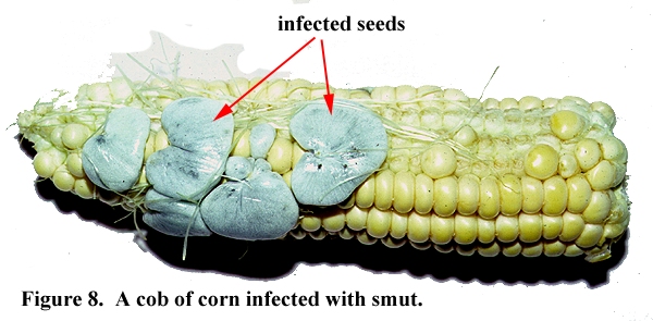
8. An infection of the maize smut fungus, Ustilago maydis, on a cob of corn. The infected seeds are inflated and greyish blue. When they are mature, the seed wall becomes brittle and breaks to release clouds of powdery spores.
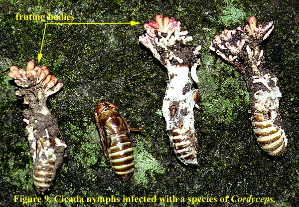
9. Cicada nymphs infected with an undescribed species of Cordyceps. The fungus has produced numbers of reddish club-shaped fruiting bodies from the top of each nymph while it was buried in the soil. Only the small, 13 mm long clubs appear on the soil surface. The interior of each nymph is a mass of white mycelium.

Many species of fungi enter a type of partnership with the roots of trees where both the fungus and the tree benefit from the association. A fungus/tree root partnership is called a mycorrhiza (a name which means 'fungus root') and a mycorrhizal internal structure can only be seen with a microscope.
Mycorrhizal relationships are sometimes very specialised, and often only very specific fungal species can enter the roots of a particular plant. Sometimes only one fungus species can form the partnership. Trees of the genus Pinus (including the economically important slash pines and radiata pines) must have their own special mycorrhizal fungi imported with them in order for the pines to grow. Australian eucalypts have similar mycorrhizal requirements if they are to grow properly in overseas locations (See figures attached to this text).
There are two basic types of mycorrhiza but only one is dealt with here as it is the type formed by macrofungi. In an ectomycorrhiza the fungus forms a coating or sheath around the tree root. Once the fungus has infected the root, it moves inside the root tissues but does not actually invade any of the root cells. Instead it squeezes between them, and when a section of the root is taken the cells of the fungus can be clearly seen between the cells of the root (See the figure attached to this text. The fungal cells are coloured purple and can easily be seen as a sheath around the root and penetrating between the root cells.).
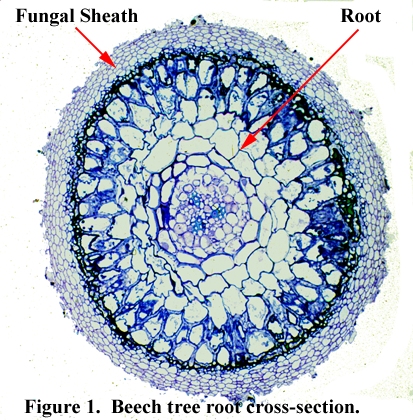
Fig 1. A cross-section of a beech tree root (Fagus sylvatica) showing the mycorrhizal sheath around the outside of the root.
The fungus can grow much faster than the roots or their root hairs and so, when the soil conditions are good, it quickly extends its hyphae out into the soil and collects water and mineral nutrients which it sends back to its cells inside the root. The root cells now take some of the water and minerals from the fungus cells and use them for the growth of the tree. Since the fungus can respond to favourable conditions very quickly and can rapidly spread through a large volume of soil, it can absorb sufficient water and minerals for good plant growth even in very poor soils. This means that trees with mycorrhizal partners are able to grow quite well in very low nutrient conditions such as the impoverished, sandy soils of the Blue Mountains of New South Wales.
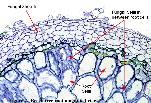
Fig 2. A magnified cross-section of a beech tree root (Fagus sylvatica) showing the fungal sheath and the fungal cells between the root cells (called the Hartig Net).
The relationship is not just one way and the fungus also benefits. If the soil becomes extremely dry, the fungus dies off, but it survives in the shelter of the root interior. In addition, the fungus takes a small amount of the sugars that have been produced in the leaves of the canopy and transported to the roots.
Many Australian plant species, including all eucalypts, form mycorrhizal relationships of the type described above. Mycorrhizae are also found on species of Tristania, Lophostemon, Melaleuca, Leptospermum and Kunzea, as well as species from different plant families, e.g. Casuarina.
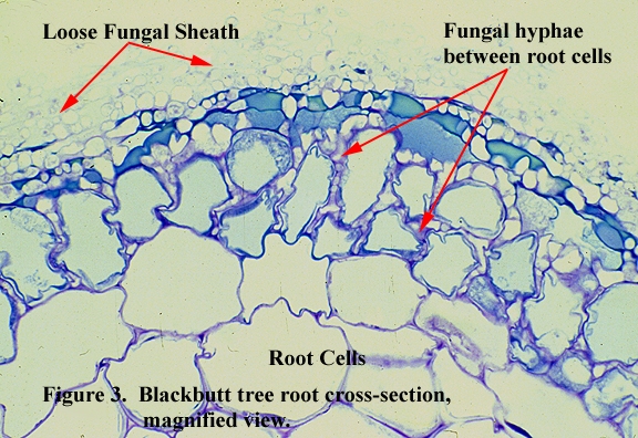
Fig 3. A magnified cross-section of a blackbutt root (Eucalyptus pilularis) showing the loose fungal sheath and the fungal cells between the root cells.
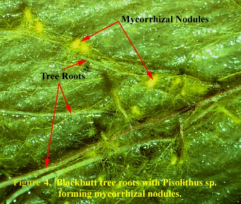
Fig 4. A species of the fungus Pisolithus has formed small, yellow nodules around the sites where it is invading the roots of blackbutt (Eucalyptus pilularis).