Glossary
| A | B | C | D | E | F | G | H | I | J | K | L | M | N | O | P | Q | R | S | T | U | V | W | X | Y | Z |
Image Gallery:
- Morphology of a teleost larva
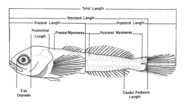
Diagrammatic representation of morphology of a teleost larva.
Auer, N. A. (ed.). 1982, Identification of Larval Fishes of the Great Lakes Basin with Emphasis on the Lake Michigan Drainage.
Great Lakes Fishery Commission, Ann Arbor, MI 48105. Special Pub. 82-3, p. 8. - Measurement locations for notochord and head depth/width
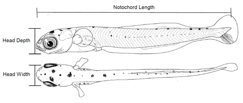
Illustration of the measurement locations for notochord length and head depth and width.
Tenera Environmental. Length-Specific Probabilities of Screen Entrainment of Larval Fishes
Based on Head Capsule Measurements, San Luis Obisbo, 2013 [using diagram from Moser, CALCOFI, "jacksmelt"] - YSL & Larva diagnostic features
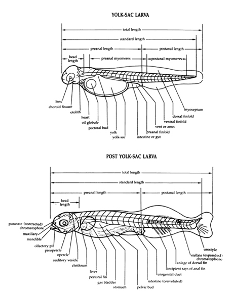
Diagrammatic representation of typical yolk-sac larva and post yolk-sac larva (redrawn from original drawing by Alice J. Mansueti in Mansueti and Hardy 1967).
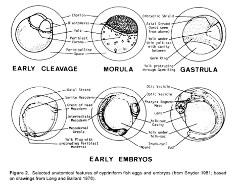
Wallus, R., T. P. Simon, and B. L. Yeager. 1990. Reproductive biology and early life history of fishes in the Ohio River drainage. Volume 1: Acipenseridae through Esocidae. Tennessee Valley Authority, Chattanooga, Tennessee, USA. p.8. - Embryo features
- Rock Bass larva with features
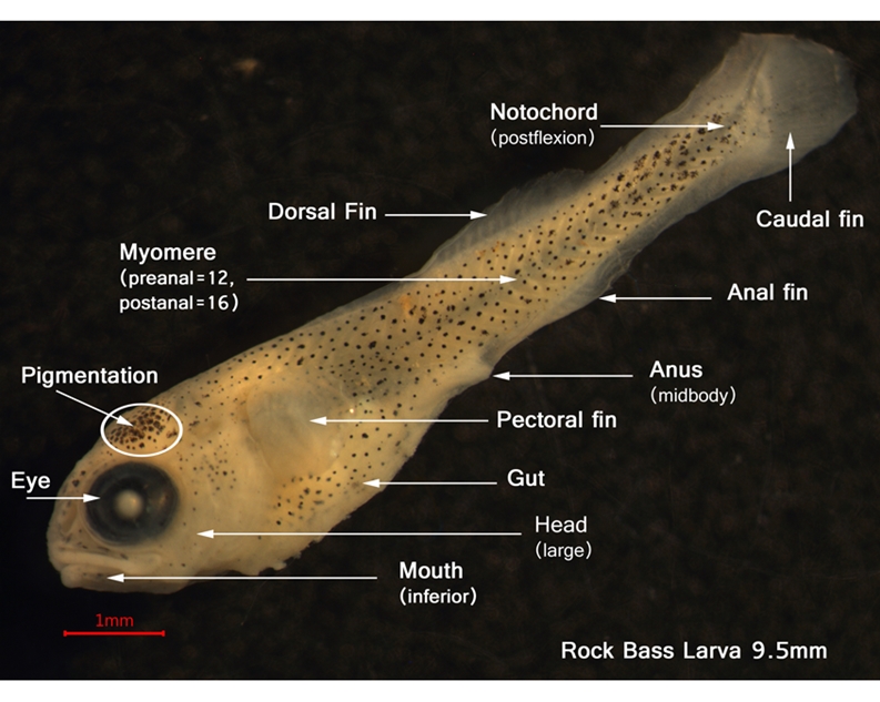
Photo courtesy of USGS Great Lakes Science Center, 2011
- Walleye YSL with features
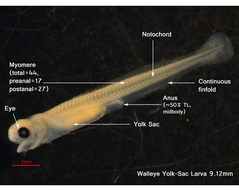
Photo courtesy of Stacey Ireland, USGS Great Lakes Science Center, 2012
A
- abbreviation for anal fin
- Abdominal
- gut region
- Abbreviate heterocercal
- tail in which the vertebral axis is prominently flexed upward, only partly invading upper lobe of caudal fin; fin fairly symmetrical externally
- Actinotrichia
- horny fin supports (threadlike fibers) which are the precursors of fin rays or spines (lepidotrichia)
- Adherent
- attached or joined together, at least at one point
- Adhesive (egg)
- referring to eggs, those which stick to each other or to a substrate after water hardening, unless otherwise noted; adhesiveness of entire egg capsule may or may not persist after attachment
- Adhesive organ
- a suctorial organ, located anteriorly on snout, which produces a viscid secretion allowing larva to attach to objects
- Adipose fin
- fleshy, rayless fin posterior to (behind) the dorsal fin (between dorsal and caudal fins)
- Adnasal pore
- pore at the end of the short, ascending branch of the infraorbital canal, which lies just behind the anterior nostril; not always present (eels)
- Adnate
- joined to; grown together
- Adnexed
- flaglike
- Adult
- sexually mature as indicated by production of gametes
- Air bladder
- see: swim bladder
- Alevin
- a term applied to juvenile catfish, trout, and salmon after yolk absorption; exhibiting no post yolk-sac larval phase
- Aliform
- wing-like; usually referring to the shape of the pectoral fin
- Allopatric
- having separate and mutually exclusive areas of geographic distribution
- Anadromous
- referring to fishes that ascend rivers to spawn
- Anal
- pertaining to the anus or vent
- Anal fin
- fin (usually single but double in some gadiforms) on the ventral margin of the tail; unpaired median fin immediately behind anus or vent
- Anal fin origin
- anterior-most point at which the anal fin attaches to the body
- Angle of jaw
- bony prominence posterior to gape of jaw; region of the junction of the angular, articular, and quadrate bones
- Angular
- the bone at the angle of the jaw or that region; used in reference to pigmentation at, or near, the angular bone
- Anlage(n)
- rudimentary form of an anatomical structure or organ; primordium
- Anterior
- towards the front; cephalad
- Antero-hyal
- anterior bone to which branchiostegal rays attach: formerly ceratohyal
- Antrorse
- angled forward; usually referring to direction of a spine
- Anus
- orifice and surrounding tissue at the terminus of gut; external orifice of the intestine; vent
- Aorta
- main blood vessel lying below the spinal chord that supplies blood from the heart
- Articular
- a bone of the lower jaw between the dentary and angular bones (some consider it part of the angular); used in reference to pigment anterior to the angle of the upper jaw
- Auditory vesicle
- sensory anlage from which the ear develops; clearly visible during early development
- Axillary process
- enlarged accessory scale attached to the upper or anterior base of pectoral or pelvic fins
Top
B
- BDA
- body depth at anus
- BL
- body length
- Band
- a strip of pigment that contrasts with the adjacent background pigment or unpigmented area; it may be on any part of the head, body, or fins and may be oriented in any position; a vertical band occurring laterally on the head or body is referred to as a bar and a horizontal band is referred to as a stripe
- Bar
- a vertical band of pigment on the lateral surface of the head or body; often occurring in a series
- Barbel
- slender sensory projection on the lips or chin; barbel length measured from the point of attachment on the head to the tip of the barbel
- Basibranchials
- three median bones on the floor of the gill chamber, joined to the ventral ends of the five gill arches
- Basin
- a large complex of rivers or lakes with a single outlet to the ocean (e.g., Great Lakes)
- Basipterygium
- bone or process that supports the pelvic fin
- Bathypelagic
- zone below 1,000m depth in the open ocean
- Benthic
- living on or in the bottom (substrate)
- Bicuspid
- having or ending in two points; a tooth with two points
- Blastocoel
- cavity of the blastula; segmentation cavity
- Blastoderm
- specifically, early embryonic tissue composed of blastomeres; more generally, embryonic tissue prior to formation of embryonic axis
- Blastodisc
- early embryo of teleosts consisting of a disc-or cap-like mass of cells on the yolk; embryo-forming area of egg prior to cleavage
- Blastomere
- individual cells forming the early embryo of teleosts (during cleavage)
- Blastopore
- opening formed by and bordered by the germ ring as it extends over the yolk
- Blastula
- stage in embryonic development which represents the final product of cleavage stages, characterized by formation of the blastocoel
- Body depth
- the vertical distance from the dorsal margin of the body to the ventral margin of the body measured at the base of the pectoral fin where it attaches to the body; fins or fin bases are not included in the measurement
- Body depth at anus
- vertical distance of body at anus
- Body length
- general term used to indicate the size of a larva; equivalent to notochord length (distance from the tip of the snout to the tip of the notochord) in preflexion and flexion stage larvae and equivalent to standard length (distance from the tip of the snout to the posterior margin of the hypural plate) in postflexion larvae. In beloniform fishes, body length measured from the end of the opercle to the caudal base because of allometry and because the beak is often broken
- Branched ray
- soft ray with two or more branches distally
- Branchial arches
- bony or cartilaginous structures, supporting the gills, filaments and rakers
- Branchial pores
- pores in branchial region of lateral-line canal (eels)
- Branchial region
- in petromyzontids, area between the anterior margin of the first gill opening and the posterior margin of the last
- Branchiostegal membrane
- membrane connecting the elongated bones which ventrally support the gill membranes
- Branchiostegal rays (branchiostegals)
- bony rays supporting the membranes which close the gill (branchial) cavity under the head; ray-like bony elements attached to the hyoid arch, extending under the gill openings and connected by a membrane
- Bud
- the undifferentiated bump or protuberance that appears at the initial formation of the paired fins
- Buoyant egg
- an egg which floats free within the water column; pelagic
Top
C
- abbreviation for caudal fin
- CPD
- caudal peduncle depth
- Caeca
- finger-like outpouchings at boundary of stomach and intestine
- Calcareous
- composed of, containing, or characteristic of calcium carbonate
- Cardiform
- brush-like; referring to teeth of uniform length in patches or bands
- Caruncles
- fleshy outgrowths; modified dorsal fin rays in ceratiids
- Catadromous
- fishes which go to sea from rivers to spawn
- Caudal fin
- median fin at the posterior end of the fish; tail fin
- Caudal peduncle
- narrow part of the tail between the posterior end of the anal or dorsal fin and the base of the caudal fin; caudal peduncle length measured from the insertion of the posterior-most ray of the anal or dorsal fin (whichever is most posteriad) to the insertion of the anterior-most caudal fin-ray
- Caudal vertebrae
- the posterior group of vertebrae extending from the centrum bearing the first haemal spine to the urostyle
- Cement glands
- discrete or diffuse structures which permit a larva to adhere to a substrate
- Centrum
- the body of a vertebra
- Cephalic
- belonging to the head
- Cerotohyal
- see: antero-hyal
- Cheek
- lateral surface of head between eye and opercle, usually excluding preopercle
- Chevron-shaped
- the earliest developmental form of myomeres in larvae; describing the angle formed by the epaxial and hypaxial portions of the myosepta
- Chorion
- after water hardening, the outermost membrane of a fish egg
- Choroid fissure
- a cleft in outer layers of the eye visible in early larvae; indentation at the ventral margin of the eye marking the invaginated borders of the optic cup in larval fish; apparent in young fish as a trough-like area below lens
- Choroid tissue
- mass of primordial vascular tissue of various shapes lying below the eye; usually associated with narrow eyes; often pigmented; its length measured along its longitudinal axis from the interface with pigmented portion of the eye to the tip of the choroid mass
- Chromatophore
- pigment-bearing cell; frequently capable of expansions and contractions which change their size, shape, and color
- Cirrus
- generally small, dermal, flap-like or tentacle-like process on the head or body
- Cleavage stages
- initial stages in embryonic development where divisions of blastomeres are clearly marked; usually include 1st through 6th cleavages (2-64 cells)
- Cleithral symphysis or junction
- where the ventral ends of the cleithral bones meet
- Cleithrum
- large bone of support for the pectoral fins; elongate vertical bone in pectoral girdle at the junction of the head and body of the fish; one of the first bones to form in the body; demarcates the junction of the head and body; clearly visible in many fish larvae
- Coelomic
- belonging to the body cavity
- Coiled
- condition of the gut where it is twisted or convoluted
- Compressed
- laterally flattened
- Confluent
- coming together to form one
- Crown
- top of the head
- Ctenoid scale
- scales having small, needle-like projections on the posterior margin; bearing cteni
- Cycloid scale
- scales with evenly curved free border, without cteni
Top
D
- D or DF
- dorsal fin
- DFO
- dorsal fin origin
- Dash (pigment)
- an elongate or streak-like melanophore
- Deciduous
- referring to scales that are easily rubbed off and thus not firmly attached
- Demersal (egg)
- referring to an egg which rests upon the substrate as a result of deposition or settling; at the ocean bottom; used in reference to life stages from eggs to adults; may be free or attached
- Dendritic
- highly branched; usually used in reference to a melanophore
- Dentary
- major bony element of the lower jaw, usually bearing teeth
- Depth
- greatest depth of body, excluding median fins (taken just behind gill opening, at or behind anus, or elsewhere at greatest depth)
- Disk
- flat cup-like structure formed from modified pelvic fin rays (also pectoral fin rays in some fishes) used for holding onto the substrate or structures on the substrate (e.g., gobiesocids, cyclopterids, gobioids); its diameter (P2DL) is measured longitudinally from one margin to the opposite margin; in echeneids, the disk is on the top of the head and formed from modified dorsal fin spines; also refers to the disk-shaped head of ogcocephalids where disk width (DW) is the greatest transverse dimension of the disk
- Distal
- remote from the point of attachment; opposite to proximal
- Distribution
- occurrence of species in the region (e.g., Great Lakes region and within the Lake Michigan drainage)
- Dorsal
- upper part of body; opposite of ventral
- Dorsal fin
- fin or fins on dorsal margin of the body; median, longitudinal, vertical fins located on the back
- Dorsal fin origin
- point where first dorsal ray or spine attaches to body
- Dorsum
- uppermost part of body; dorsal body margin; opposite to ventrum
- Drainage
- a group of lakes or streams within a basin (e.g., Lake Michigan drainage)
Top
E
- ED
- eye diameter
- Early embryo
- stage in embryonic development characterized by formation of embryonic axis
- Early life history
- the early phase in life, spanning the developmental stages from egg to juvenile
- Egg capsule
- outermost encapsulating structure of the egg, consisting of one or more membranes; the protective shell
- Egg diameter
- in nearly spherical eggs, greatest diameter; in elliptical eggs given as two measurements, the greatest diameter or major axis and the least diameter or minor axis
- Egg pit
- the pit or pocket in a redd (nest) into which a trout female deposits one batch of eggs
- Eggs
- description of fertilized, water hardened eggs, unless otherwise noted; diameter, color, adhesive properties, buoyancy, yolk and oil globule characteristics and incubation period
- Elver
- stage of eels after glass eel stage and prior to adulthood, when they become pigmented
- Emarginate
- caudal fin possessing a slight notch or indentation, but not definitely forked
- Embryo
- organism at an early stage of differentiation and growth; refers to the stage of development between fertilization and hatching
- Embryonic axis
- primitive differentiation of the embryo; an elongate thickening of blastodermal tissue
- Embryonic shield
- thickened area of the germ ring representing the future longitudinal axis of the embryo
- Emergence
- the act of leaving the substrate and beginning to swim; swim-up
- Engyodontic stage
- early stage of anguilliform larvae (preceding euryodontic stage) characterized by few needle-like teeth, the upper and lower jaws equal in length, no nasal capsule, an undifferentiated finfold, no hypurals, the notochord tip straight, and the head and preanal region of the body relatively large
- Epaxial
- the portion of the body above the horizontal myoseptum; the part of the myomeres above the lateral midline (horizontal septum)
- Epibenthic
- zone at the interface with the sea bottom; refers to organisms living in contact with the sea bottom
- Epiboly
- movement of embryonic cell mass over the surface of the yolk; the germ ring marks the boundary of the advancing sheet of cells
- Epihyal
- see: postero-hyal
- Epipelagic
- zone from the surface to 200m depth in the open ocean
- Epurals
- modified vertebral elements which lay above the vertebrae and support part of the caudal fin
- Erythrophores
- red or orange pigment cells
- Esca
- the lure at the tip of illicium in most lophiiform fishes; commonly luminous in deepsea species
- Esophagus
- alimentary tract between pharynx and stomach
- Ethmoid pore
- pore in the ethmoid canal, included in the count for supraorbital pores (eels)
- Euryodontic stage
- advanced stage of anguilliform larvae (following engyodontic stage) characterized by a relatively smaller head and preanal region, three series of relatively short, broad teeth, a relatively shortened lower jaw, the formation of nasal capsules, fins, and hypurals, and the flexion of the notochord
- Eye diameter
- horizontal distance of the iris of the eye; in larvae with round eyes, the diameter of the pigmented part of the eye, usually measured through the horizontal midline; in larvae with oval or elliptical eyes, the horizontal dimension is given first, followed by the vertical dimension
- Eye length
- the longer axis of an oval or elliptical eye measured through the midline from the pigmented margin of one side of the eye to the other side
- Eye stalks
- movable peduncles of varying length bearing the eyes; eye stalk length measured from the point where the stalk attaches to the head to the point of attachment at the eye
- Eye width
- the shorter axis of an oval or elliptical eye measured through the midline from the pigmented margin of one side of the eye to the other side
Top
F
- FL
- fork length
- Falcate
- scythe-shaped; referring to an anal fin; deeply concave as a fin with middle rays much shorter than anterior and posterior rays
- Fecundity
- average number or range of ovarian eggs per female, unless otherwise noted
- Fin elements
- fin spines, rays, and supporting bones (pterygiophores)
- Fin insertion
- posterior-most point at which the fin attaches to the body
- Fin origin
- anterior-most point at which the fin attaches to the body
- Fin rays
- segmented cartilage or bony structures which support fin membranes
- Finfold
- median fold of skin surrounding the body within which the dorsal, anal, and caudal fins develop; the part of the ventral finfold in which the anal fin develops is referred to as the anal finfold
- Finlet
- small fin-like structures posterior to the dorsal and anal fins
- First-feeding larvae
- larvae that have used all or most of their yolk and are capable of capturing prey
- Flexion
- stage (or the process) when the urostyle bends dorsally concurrently with the development of principal rays and hypural bones in the caudal fin; also, the process of notochord flexion; preflexion refers to the stage prior to the initial bending of the notochord tip and postflexion refers to the stage after the completion of the notochord flexion
- Focal point
- location of a fish maintaining a stationary position on or off the substrate for at least a 10-second period
- Fontinelle
- a gap or space between bones in the roof of the skull covered only by a membrane
- Foramen
- an opening through a bone
- Forebrain
- anterior region of the developing brain that includes the olfactory lobes
- Foregut
- anterior part of the primitive alimentary canal from which the esophagus and stomach develop
- Fork length (FL)
- distance from the most anterior point on the snout to the end of the shortest central caudal fin ray
- Frenum
- a fold of skin that limits movement of the upper jaw
- Frontal bones
- large paired (fused in some species) bones that form the top of the cranium anteriorly; they often from a ridge over each orbit (supraocular crest) that may bear one or more spines
- Frontal pore
- a single median pore found in the frontal canal, present in some families (eels)
Top
G
- GB
- location of the gall bladder
- GD
- greatest depth
- Ganoid scales
- diamond- or rhombic-shaped scales consisting of bone covered with enamel
- Gape
- the border of the mouth
- Gargaropteron stage
- pelagic stage of the chiasmodontid genus Kali, characterized by greatly elongate pectoral and pelvic fin rays
- Gas bladder
- see: swim bladder
- Gastrula
- stage in embryonic development between blastula and embryonic axis
- Germ ring
- thickened margin of the blastodisc that advances over the yolk during epiboly
- Germinal disc
- the blastodisc
- Gill arch(es)
- see: branchial arches
- Gill opening
- measure distance between upper and lower ends of gill opening
- Gill rakers
- variously-shaped bony projections on anterior edge of the gill arches
- Glass eel
- stage of eel following leptocephali stage when they enter estuaries (after pigmentation they are called elvers)
- Glossohyal
- a median bone of the tongue
- Granular yolk
- yolk consisting of discrete units of finely to coarsely granular material
- Greatest body depth
- greatest vertical distance of body excluding fins and/or finfolds
- Guanophores
- silvery or white pigment cells containing iridescent crystals of guanine
- Gular
- ventral region of the head anterior to the isthmus and below the lower jaw
- Gular fold
- transverse membrane across throat
- Gular plate
- median ventral bony plate or plates located behind the chin and between the sides of the lower jaw, as in Amia calva; large dermal bone on throat
- Gular region
- throat
- Gut
- alimentary tube and associated organs
- Gut length
- distance from anterior margin of yolk sac to vent or from anterior margin of stomach (when distinguishable) to vent
- Gut loop
- loop, fold, or curve found along the axis of the gut
Top
H
- HD
- head depth
- HL
- head length
- HW
- head width
- Haemal spine
- a median spine on the ventral surface of a vertebral centrum, attached to the centrum by two bones that form an arch (haemal arch)
- Head length (HL)
- distance from the most anterior point on the snout to the posterior edge of the auditory vesicle, cleithrum or opercle as each develop; horizontal distance from the tip of the snout to the posterior margin of the cleithrum; or to posterodorsal point of gill opening, depending upon family
- Head width (HW)
- greatest dimension between opercles; transverse distance between the lateral margins of the head measured at the posterior margin of the orbit
- Heterocercal
- caudal fin upper lobe much larger or longer than lower; tail in which the vertebral axis is flexed upward and extends nearly to tip of upper lobe of caudal fin; fin typically asymmetrical externally,
- Hindbrain
- posterior region of the developing brain that includes the medulla
- Hindgut
- posterior part of the alimentary canal that includes the intestine and rectum
- Holoblastic
- type of cleavage in which the entire egg, including the yolk, undergoes division
- Homocercal
- type of caudal fin in teleosts where the upper and lower lobes are symmetrical; internally, the major structural elements of the fin are an upturned urostyle (several fused vertebrae) that articulates with flattened bones (hypurals and parhypural) that support the principal fin rays
- Homogeneous
- uniform in composition; opposite to segmented in referring to egg yolk
- Horizontal myoseptum / septum
- a sheet of connective tissue (in the horizontal plane) that separates the epaxial and hypaxial muscle masses; the lateral midline area in reference to pigmentation
- Hyomandibular
- bone or cartilage (usually elongate) in the cheek region that functions in jaw suspension
- Hypaxial
- the portion of the body below the horizontal myoseptum; the part of the myomeres below the lateral midline (horizontal septum)
- Hypochord(al)
- below the notochord; referring to the lower lobe of the caudal fin; a transitional rod of cells which develops under the notochord in the trunk region of some embryos
- Hypurals
- the expanded haemal spines of the posterior vertebrae which support most of the caudal fin
Top
I
- Ichthyoplankton
- that part of the zooplankton consisting of the egg and larval stages of fishes
- Illicium
- tentacle-like spine of the dorsal fin located on the snout of most lophiiform fishes, used as a lure to attract prey; illicium length measured from the point of insertion of the illicium on the head to the tip of the structure
- Incipient
- becoming apparent
- Incubation period
- time from fertilization of egg to hatching
- Inferior
- spatial anatomical term meaning lower in position; opposite to superior
- Inferior mouth
- snout projecting beyond the lower jaw; also referred to as subterminal mouth
- Infraorbital
- space between eyes over top of head
- Infraorbital pores
- pores in infraorbital canal, including pores in lateral canal along upper jaw below eye, and postorbital pores (sometimes listed separately) in ascending canal behind eye (eels)
- Insertion (of fin)
- posterior (usually) point of attachment of a fin
- Integument
- an enveloping layer or membrane; coating or external skin
- Internarial
- area between the nares on one side of the head or the other
- Interopercle
- lower bone of the gill cover lying below the preopercle
- Interorbital
- region on top of the head between the orbits of the eyes
- Interorbital pores
- pores in the supraorbital canal which are located between the eyes (eels)
- Interorbital width
- least distance between the orbits across dorsum of head
- Interradial
- area between the fin rays
- Interspaces
- spaces between parr marks in salmonids
- Iridocytes
- crystals of guanine having reflective and iridescent qualities
- Iridophores
- see: guanophores
- Isocercal
- symmetrical tail fin; vertebral column ending along the median line, caudal fin rays arising symmetrically from it; tail in which vertebral axis terminates in median line of fin, as in Gadiformes
- Isthmus
- fleshy space beneath the head and between the gill openings; ventral region of the head below the gills, often narrow, connecting the gular and cleithral regions; measure interbranchial distance as ventral distance between lower ends of gill opening
Top
J
- Jugular
- referring to the throat region; beneath the head; usually used to indicate the position of the pelvic fin in some fishes
- Juvenile
- phase of development from complete fin ray development and finfold absorption to sexual maturity; stage after transformation from the larval stage that is like the adult but not yet reproductively active; there is a full complement of fin rays, scales are formed (in species that have them), and the form is fundamentally similar to the adult
Top
K
- Keeled
- with a ridge or ridges
- Kupffer's vesicle
- a small, vesicular, ventro-caudal pocketing which forms as blastopore narrows
Top
L
- LIV
- location of the liver
- Lachrymal
- the anterior-most of the infraorbital (circumorbital) bones; enlarged and with spines in some species
- Lanceolate
- slightly broad at the base and tapering to a point
- Larva / larvae
- phase of development from complete absorption of yolk to development of the full complement of adult fin rays and absorption of finfold; stage following hatching that is unlike the juvenile or adult in form and pigmentation, and must transform or metamorphose before assuming juvenile/adult characteristics
- Last vertical blood vessel
- the posterior-most large blood vessel extending from the dorsal aorta to the kidney (nephros); used as reference point and taxonomic character in eel leptocephali
- Late embryo
- stage prior to hatching in which the embryo has developed external characteristics of its hatching stage
- Lateral line
- a line along the lateral surface of the body (backward from head along sides) formed by a series of sensory pores that usually are associated with modified scales
- Lateral line pores
- pores in lateral-line canal (sometimes counted to anus, sometimes total count given) (eels)
- Lateral line scales
- pored or notched scales associated with the lateral line
- Lateral midline
- the region of the body between epaxial and hypaxial myomeres; the region of the horizontal septum
- Lateral series scales
- number of rows of scales crossing the midlateral surface or lateral line if complete
- Lateral teeth
- in petromyzontids, teeth of oral disc lateral to esophageal opening
- Lepidotrichia
- replacements of actinotrichia; soft fin rays or spines; scale-like structures that form the segments of soft rays in bony fishes
- Leptocephalus (leptocephali)
- transparent, ribbon-like, often large larvae of elopiform fishes with an internal cavity filled with acellular mucinous material; leptocephali usually have a small head and prominent teeth
- Lithophils
- requiring gravel as a substrate for spawning
- Live-bearing
- see: viviparous
- Longitudinal septum
- a sheet of connective tissue that separates the muscle masses on the right and left sides; the dorsal longitudinal septum separates the epaxial muscle masses and the ventral longitudinal septum separates the hypaxial masses
- Low terminal mouth
- mouth that is slightly oblique to horizontal with anterior end of upper lip at or below bottom-of-eye level and either even with or the most anterior margin of snout
- Lower jaw
- measure from tip of lower jaw to angle of mouth
- Lunate
- crescent (moon) shaped
Top
M
- mm
- millimeter
- Mandible
- lower jaw, comprised of three bones: dentary, angular and articular
- Mandibular pores
- pores along lower jaw, part of preoperculomandibular canal series (eels)
- Maxilla / maxillae
- the posterior, lateral bones of the upper jaw; the longest paired bones of the upper jaw; located above the premaxillae
- Maxillary
- the dorsalmost of the two bones in the upper jaw
- Meckel's cartilage
- embryonic cartilaginous axis of the lower jaw in bony fishes
- Median
- referring to the midline plane that divides a bilaterally symmetrical animal into right and left halves; usually synonymous with mesial
- Melanophore
- melanin-bearing pigment cell; ameboid black or brown pigment cells of various shapes and sizes derived from the neural crest region of the embryo; black chromatophores
- Mental
- pertaining to the chin
- Meristic characters / meristics
- countable structures occurring in series (e.g., myomeres, vertebrae, fin rays)
- Meroblastic
- type of cleavage in which only the blastodisc undergoes division
- Mesencephalon
- midbrain; serves optic functions
- Mesolarva
- phase of larval development (used by Snyder et. al.) characterized by presence of at least one dorsal, anal, or caudal-fin spine or ray but either lacking the adult complement of principal soft rays in at least one median (dorsal, anal, or caudal) fin or lacking pelvic-fin buds or pelvic fins (if present in adult)
- Mesopelagic
- zone from 200m to 1000m depth in the open ocean
- Mesopterygoid
- middle of three dermal bones of the upper jaw
- Metalarva
- phase of larval development (used by Snyder et. al.) characterized by presence of adult complement of principal soft rays in all median fins and pelvic-fin buds or pelvic fins (if present in adult)
- Metamorphosis
- a marked change in form or structure at the end of the larval stage involving acquisition of adult characters and loss of larval characters; synonymous with transformation
- Metencephalon
- portion of the brain immediately behind the mesencephalon
- Micropyle
- principle path of sperm entry through the chorion (vitelline membrane) of an egg
- Midbrain
- region of the developing brain that includes the optic and cerebellar lobes
- Midline
- the median plane or line of the body either on the dorsal or ventral surface; sometimes used as the middle of a body part
- Molariform
- referring to a tooth with a flat grinding surface
- Morula
- stage in development of egg in which blastomeres from a mulberry-like cluster
- Mucosal folds
- folds of tissue lining the cavity of the intestine; pronounced in some fish larvae giving a striated or rugose appearance
- Myomere formula
- preanal myomeres + postanal myomeres = total myomeres
- Myomeres
- muscle segments of the body occurring in series; approximately equal to number of vertebrae in adults; the preanal myomeres are defined as all myomeres anterior to a vertical from the posterior margin of the anus and postanal myomeres are those myomeres posterior to a vertical from the posterior margin of the anus; in many fishes the number of preanal and postanal myomeres approximate the number of precaudal (abdominal) and caudal vertebrae, respectively
- Myoseptum / myosepta
- thin partition of connective tissue which joins myomeres
Top
N
- NL
- notochord length (see also standard length - used as, prior to and during notochord flexion)
- Nape
- dorsal region of the body immediately behind the head
- Nares
- nostrils, openings leading to the olfactory organs; the openings of the nasal organs or rosettes
- Narial
- pertaining to the nares
- Nasal
- pertaining to region of the nostrils, or to the specific bone in that region
- Nasal capsule
- paired, more-or-less spherical structures on the snout that contain the olfactory organs
- Natural hybrids
- other species in the study area with which interbreeding has occurred naturally
- Nekton
- motile, marine organisms living in open water (rather than the sea floor) and capable of swimming against currents; small to moderate-sized nektonic organisms (e.g., midwater fishes, juvenile fishes, some cephalopods) are referred to as micronekton
- Nephros
- kidney; often used to refer to larval kidney
- Neritic
- pelagic coastal zone extending seaward to the margin of the continental shelf
- Neural crest
- region of the neural ridge of the developing embryo that differentiates into many kinds of tissue and cells, including melanophores
- Neural spine
- a median spine on the dorsal surface of a vertebral centrum, attached to the centrum by two bones that form an arch (neural arch) through which the spinal cord passes
- Neustonic
- inhabiting the surface of the ocean; plankton living in this zone are referred to as neuston
- Notochord
- longitudinal cartilaginous rod that supports the axis of the body; eventually replaced by the vertebral column in teleostean fishes
- Notochord length (NL)
- the distance from the tip of the snout to the posterior tip of the notochord; used prior to and during notochord flexion (see also: standard length)
- Nuchal
- referring to the region of the nape; immediately behind the head, dorsally
Top
O
- Obtuse
- with a blunt or rounded end; an angle greater than 90 degrees
- Occipital crest
- bony ridge located posteriorly on top of the head
- Occipital region
- area on dorsal surface of head, beginning above or immediately behind eyes and extending backwards to end of head
- Occiput
- see: occipital region
- Oceanic
- open sea zone seaward of the continental shelf or slope
- Oil globule
- discrete spheres of oil or fatty material within the yolk of some fish eggs
- Olfactory buds
- incipient olfactory organs
- Ontogenetic characters
- those characters associated with developmental stages
- Ontogeny
- developmental history of an organism from zygote to maturity
- Opercle
- upper posterior and usually largest bone of the gill cover of a fish; often used as synonym of operculum
- Operculum
- bony plate of the gill cover
- Ophioblennium stage
- pelagic stage of some salariin blenniids characterized by enlarged pectoral fins and enlarged, hooked teeth anteriorly in the lower jaw or in both jaws
- Opisthonephros
- the embryonic or larval kidney
- Optic vesicles
- embryonic vesicular structures which give rise to the eyes
- Orbit
- the bony socket of the eye
- Organogenesis
- the relatively advanced period of embryonic development characterized by formation of the organ systems
- Origin (of fin)
- anterior (usually) point of attachment of a fin
- Ossification
- process of bone formation involving calcification of cartilage or connective tissue
- Otic
- region of the head containing the auditory or hearing organs
- Otoliths
- small, calcareous, secreted bodies within the inner ear
- Over yearling
- fish having spent at least one winter in a stream; applies to trout and salmon
- Oviparous
- producing eggs that develop outside the maternal body
Top
P
- P or P1
- pectoral fin
- P2 or V
- pelvic fin
- PAL
- preanal length
- PosAL
- postanal length
- PreAL
- preanal length
- PreDFL
- predorsal fin length
- PreDFFL
- predorsal finfold length
- Paedomorphic
- referring to the phylogenetic retention of juvenile (or larval) characters in the adult stage
- Palatine teeth
- teeth on the paired palatine bones in the roof of the mouth of some fishes
- Palatines
- paired bones on the roof of the mouth, often bearing teeth
- Papilla
- a fleshy projection or protuberance
- Parapatric
- distribution of species or other taxa that meet in a very narrow zone of overlap
- Paravertebral
- along the same plane as the spinal column
- Parhypural
- lowermost supporting bone of the principal caudal fin rays
- Parietal
- paired bones of the roof of the skull
- Parr marks
- dark vertical oval bars along the sides, typical of salmonids
- Pectoral (fin) bud
- swelling at site of future pectoral fin; anlage of pectoral fin
- Pectoral fin
- paired lateral (sometimes ventrolateral) fins behind the head, articulating with the pectoral girdle
- Pectoral fin base
- supporting structure of the pectoral fin; in larvae it is peduncular and contains the muscles that operate the fin; its length is measured along the longitudinal axis from the point of insertion on the body to the point of attachment of the fin blade or rays; its depth is measured on the transverse axis at its widest (usually most distal) point
- Pectoral fin length
- distance from base to farthest tip of fin; prior to ray formation, pectoral fin length is measured from the base of the blade (finfold) to the greatest distal margin of the blade; after ray formation, pectoral fin length is measured from the point of insertion of the longest ray to the tip of the ray
- Pedicel
- a small, short stalk
- Peduncle
- fleshy end of the body between the anal and caudal fins; a narrow part or stalk that connects a structure to the body (e.g., caudal peduncle connecting caudal fin to body)
- Pelagic
- living in the open water habitat, as opposed to bottom living or inshore inhabitants; free-living in the sea away from the sea bottom, usually beyond the continental shelf; not necessarily near the surface
- Pelvic bud
- swelling at site of future pelvic (ventral) fins; anlage of pelvic fin
- Pelvic fins
- paired fins usually located ventrally on the body; various in position from beneath the head (jugular), to below the pectoral region (thoracic), to the gut region (abdominal); high on the sides in some stomioids; prior to ray formation, pelvic fin length is measured from the base of the blade (finfold) to the greatest distal margin of the blade; after ray formation pelvic fin length is measured from the point of insertion of the longest ray to the tip of the ray; articulating with the pelvic girdle; ventral fins
- Periblast
- thin membrane lying below the embryo and surrounding the yolk in teleosts; the space surrounding the yolk is invaded by nuclei, forming a syncytial region of unknown function; observed as a thin border around blastula
- Pericardium
- cavity in which the heart lies
- Peritoneal
- region of the body associated with the gut or the membrane of the peritoneum; often synonymous with perivisceral
- Peritoneum
- membranous lining of the abdominal cavity; the membrane and associated connective tissue lining the gut cavity
- Perivitelline space
- fluid-filled space between the embryo and shell or chorion and yolk material of an egg. Width of perivitelline space is the distance between yolk and egg capsule expressed either as direct measurement or a ratio of the egg diameter
- Pharyngeal teeth
- bony tooth-like projections derived from the fifth (
- Pharyngeal) gill arch; teeth on the pharyngeal bones of the branchial skeleton
- Photophores
- luminous organs
- Phylogenetic
- referring to the evolutionary lineage of an organism
- Physoclistic
- having no connection between the esophagus and the pneumatic duct; typical of perciform fishes
- Physostomous
- having the swim bladder connected to the esophagus by the pneumatic duct
- Plankton
- small, free-living, weakly swimming or passively floating marine or fresh water organisms that drift with the currents
- Plicae
- wrinkle-like folds found on the lips of some catostomids
- Post yolk-sac larva(e)
- see: larva
- Postanal
- posterior to the anus
- Postanal length (PosAL)
- distance from the most posterior point of the anus to the most posterior point on the caudal fin or median finfold
- Postanal myomeres
- number of whole myomeres posterior to an imaginary vertical line at the most posterior point of the anus, including one urostylar element
- Posterior
- towards the back or caudal region; opposite to anterior
- Postero-hyal
- posterior bone to which branchiostegal rays attach, formerly epihyal
- Postflexion larva
- phase following upward flexion of the tip of the notochord (more precisely considered to begin with formation of all principal caudal fin rays) [note: for purposes of these keys, we do not make a distinction between pre- and post-flexion stages]
- Postorbital
- behind the eye or eye socket
- Postorbital length
- distance from posterior margin of eye to posterior edge of opercular membrane
- Postorbital pores
- pores in ascending canal behind eye, part of infraorbital canal series (eels)
- Posttemporal spine
- a spine that emerges from the posttemporal bone located on the posterolateral upper region of the skull
- Preanal
- located anterior to the anus
- Preanal length (PAL or PreAL)
- distance from the most anterior point on the snout to the most posterior point of the anus; synonymous with snout-anus distance
- Preanal myomeres
- number of myomeres from the nape to, and including any myomeres bisected by, an imaginary vertical line at the most posterior point of the anus
- Preanal vertebrae
- from the first vertebra to and including the vertebra intersected by a line drawn perpendicularly from the base of the first anal-fin ray (eels)
- Prebranchial length
- in petromyzontids, distance between the tip of the snout and the anterior margin of the first gill opening
- Precaudal vertebrae
- the anterior group of vertebrae that includes all centra anterior to the centrum with the first heamal spine
- Precocious
- specialized early formation of a structure (e.g., fins or fin elements) compared to typical developmental sequences in most fishes; does not infer abnormality
- Predorsal length
- distance from the most anterior point on the snout to the anterior margin of the base of the first dorsal fin ray when formed
- Predorsal myomeres
- number of myomeres from nape to dorsal origin of median finfold
- Predorsal scales
- scales along dorsal ridge from occiput to origin of dorsal fin
- Predorsal vertebrae
- from the first vertebra to and including the vertebra intersected by a line drawn perpendicularly from the base of the first dorsal-fin ray (eels)
- Preflexion larva
- phase between hatching and upward flexing of the tip of the notochord (or appearance of first caudal fin rays] [note: for purposes of these keys, we do not make a distinction between pre- and post-flexion stages]
- Prejuvenile
- developmental stage immediately following acquisition of minimum fin ray complement of adult and before assumption of adult-like body form; used only where strikingly different from juvenile
- Premaxilla / premaxillae
- primary bone of the upper jaw in most fish, usually bearing teeth; paired bones of the upper jaw anterior to the maxillae and usually bearing teeth; often protrusile, and extending ventrad of most of the maxillae in advanced teleosts
- Premaxillary
- the ventralmost of the two bones included in the upper jaw
- Preopercle
- upper anterior bone of the gill cover, often bearing serial spines; spines at the margin of the bone are referred to as posterior preopercular spines and those on a bony ridge forward of the margin are referred to as anterior preopercular spines
- Preoperculomandibular pores
- pores in preoperculomandibular canal, including mandibular pores along lower jaw and those in preopercular canal (both sometimes listed separately) (eels)
- Preorbital
- large bone anterior to the eye
- Primordium
- rudimentary form of an anatomical structure; anlage
- Principle anal and dorsal fin rays
- in certain fishes (e.g., Cyprinidae and Catostomidae), the principal rays include the branched rays plus one unbranched ray [the anteriorly adjacent, usually longest, unbranched ray]; the last two bases [branched rays, both of which articulate with the most posterior pterygiophore] are counted as one ray.
- Principle caudal rays
- caudal-fin rays originating on the hypural and parhypural elements; the number of principal rays is generally defined as the number of branched rays plus two.
- Procurrent caudal rays
- small dorsal and ventral rays of the caudal fin located anterior to the principal rays and not supported by hypural/parhypural elements
- Pronephric ducts
- ducts of pronephric kidney of early developmental stages
- Protocercal tail
- form with body axis extending through the caudal finfold
- Protolarva
- phase of larval development (used by Snyder et. al.) characterized by absence of dorsal, anal, and caudal fin spines and rays
- Protractile
- describing premaxillae which can be extended
- Proximal
- near the point of attachment or origin; opposite to distal
- Psammophils
- requiring sand as a substrate for spawning
- Pterotic spines
- spines emerging from the pterotic bone located posterior to the upper region of the orbit in the temporal region of the skull
- Pterygoid
- dermal bone of the upper jaw
- Pterygiophore
- bone of the anal internal skeleton supporting the dorsal and fins; cartilaginous or bony elements that form the fin base and support the fin rays of a fish
- Punctate melanophore
- round or dot-like melanophore
Top
Q
Top
R
- Rays
- segmented fin supports that are usually bilaterally paired and often branched
- Redd
- an excavated area or nest into which trout spawn
- Reticulated
- net-like or web-like in appearance
- Retrorse
- angled backward; usually referring to direction of a spine
- River system
- a group of streams or lakes which lead into a drainage (e.g., Fox
- River system)
- Rostrum
- a prolongation of the snout, sometimes ending in a spine (rostral spine); rostrum length measured from the anterior edge of the upper jaw to the tip of the rostrum or spine (holocentrids)
- Rudimentary fin rays
- in certain fishes (e.g., Cyprinidae and Catostomidae), size-graded series of shorter, unbranched soft rays anterior to the principal rays of the dorsal, anal, and caudal fins; also called secondary rays or, in the case of the caudal fin, procurrent rays; In traditional texts, formulas are represented by lower case
- Roman numerals
- , e.g., iv instead of 4 and separated from the principal ray count in Arabic, by commas. These are not able to be displayed appropriately in an electronic key.
- Rugose
- having a wrinkled appearance; often used to describe bone with a highly textured surface; sometimes used to describe the appearance of the gut, caused by numerous mucosal folds
Top
S
- SL
- standard length
- Sn-A
- snout to anus
- Saddle markings
- pigment patterns which cover the dorsal and lateral aspects and give an overall appearance of a saddle
- Sculptured
- referring to an egg shell with ornamentation or surface features of various shapes and textures
- Scute
- a modified, thickened scale, often spiny or keeled
- Secondary fin rays
- see: rudimentary fin rays or procurrent rays
- Segmented
- particulate or divided; opposite of homogeneous in referring to egg yolk
- Semibuoyant
- referring to eggs which do not float nor sink, but remain suspended in the water column
- Shell
- the membrane that encloses an egg; generally, equivalent to chorion
- Sigmoid heart
- the
- S-shaped heart which develops from the primitive heart tube
- Snout
- forward part of the head anterior to the eye; snout length measured in the longitudinal axis from the anteriormost pigmented surface of the eye to the tip of the snout
- Snout-anus (Sn-A) (or snout-to-vent) length
- distance from the tip of the snout to the posterior margin of the anus, measured at the longitudinal axis; equivalent to preanal length (PAL)
- Snout to rictus (upper jaw)
- tip of snout to angle of mouth
- Soft rays
- bilaterally paired, usually segmented, fin supports
- Somites
- primitive, segmented, mesodermal tissue along each side of notochord
- Spatulate
- having a rounded apex and tapering to a base; flattened or spoon-shaped
- Spawning habitat
- description of the environment in which spawning has been documented
- Spawning season
- months during which spawning has been documented for the Great Lakes region, unless otherwise stated
- Spawning substrate
- type of material upon which spawning is known to occur
- Spawning temperature
- water temperatures at which spawning has been documented
- Speleophils
- requiring cavities as a substrate for spawning
- Sphenotic
- bone at the upper part of the skull, often forming part of the orbit
- Spines
- supporting elements in the fins that are unsegmented, unpaired, unbranched, and usually (but not always) stiff, pungent and sharp (sometimes referred to as spinous rays); also, refers to pointed projections arising from various bones, usually on the head
- Spinous rays
- in certain otherwise soft-rayed fish, soft rays that during embryonic or larval development are thickened, fused, and hardened into spine-like structures, sometimes with moderate to strong serrations or barbs along their posterior margins. In formulas for fin-ray counts, fully spinous rays may be designated by Roman numerals like true spines.
- Spinous scale
- specialized larval scales with spines (not the serrated ctenoid scale of adult fishes)
- Squamation
- covering of scales
- Stalked eye
- eye borne on a stalk or peduncle
- Standard length (SL)
- distance from the most anterior point on the snout to the most posterior point of the notochord or hypural complex
- Stellate melanophore
- referring to a melanophore which is expanded into a star-like shape; the stellate condition can be temporary and the melanophore can become punctate after contraction
- Stomodeum
- primordial mouth; the anterior pitted portion of the embryonic gut
- Striations
- surface features in the form of lines or bands
- Stripe
- a horizontal band of pigment on the lateral surface of the head or body; sometimes occurring in a series
- Subcutaneous
- occurring beneath the skin
- Submandibular
- beneath the lower jaw; along the edge of the lower jaw
- Subopercle
- posterior bone of the gill cover lying below the opercle
- Subterminal mouth
- underneath or set back from the tip of the snout, sometimes referred to as an inferior mouth
- Superior
- spatial anatomical term meaning upper in position; opposite to inferior
- Superior mouth
- condition when the lower jaw extends upward and the mouth opens dorsally
- Supracleithral spine
- a spine originating from the supracleithrum located near the upper posterior margin of the head
- Supramaxilla
- small dermal bone attached posterior and dorsal to the maxilla
- Supraoccipital spine
- spine or crest on the midline of the back of the head originating from the supraoccipital bone
- Supraoral
- above the mouth; referring to the teeth of the oral disc in lampreys which are anterior to the mouth opening
- Supraoral tooth plate
- in petromyzontids, tooth plate immediately anterior to esophageal opening
- Supraorbital pores
- pores on dorsal surface of snout and head, including those in ethmoid canal at tip of snout (sometimes listed separately) and those on dorsal surface of snout and head; the posteriormost pores situated between the eyes are sometimes referred to as interorbital pores (eels)
- Supratemporal pores total number of pores in supratemporal canal (usually including one median pore) (eels)
- Swim bladder
- gas-filled sac lying beneath the spinal column in the abdominal region (between the kidneys and alimentary canal in teleosts); also referred to as air bladder or gas bladder
- Sympatric
- species inhabiting the same or overlapping geographic areas
- Syncytium
- a multinucleate mass of protoplasm
Top
T
- TL
- total length
- Tail
- portion of the body posterior to the anus; the postanal region
- Tail-bud stage
- stage of embryonic development characterized by a prominent caudal bulge and marked development of cephalic region
- Tail-free stage
- stage of embryonic development characterized by separation of the tail from the yolk
- Tail length
- anus to tail tip; in petromyzontids, distance from cloacal slit to tip of caudal fin
- Taxon / Taxa
- taxonomic category or unit, as a species, genus, family, etc.
- Teleosts
- bony fishes
- Telescopic eye
- type of elongate, cylindrical eye that protrudes forward or upward within an envelope of skin
- Temporal canal
- connects the lateral line with the postorbital portion of the infraorbital canal; rarely contains pores (eels)
- Terminal mouth
- condition when lower and upper jaws are equal in length and the mouth opens terminally; type of mouth that opens anteriorly; typical of most fish larvae
- Tesselated
- markings or colors arranged into squares
- Thoracic
- pelvic region; referring to the chest area; usually the region of the body of a fish below the pectoral fin base
- Total length (TL)
- distance from the most anterior point on the snout to the most posterior point on the caudal fin or fin fold
- Total vertebrae
- total number of vertebrae including the first reduced vertebra and the hypural (eels)
- Transformation
- the process (synonymous with metamorphosis) at the end of the larval stage, characterized by a marked change in form or structure and involving acquisition of juvenile or adult characters and loss of larval characters; also refers to the stage where this process occurs; the term "transitional" is used sometimes for larvae that undergo a gradual transformation (e.g., in scombrids)
- Truncate
- ending abruptly along a vertical line
- Trunk
- portion of the body between the head and the anus; from posterodorsal point of gill opening to anus
- Trunk length
- in petromyzontids, distance between posterior margin of last gill opening and cloacal slit
- Trunk myomeres
- in petromyzontids, myomeres between the most posterior gill opening and the cloacal slit
- Tubercle
- small knobby protuberance
Top
U
- Urohyal
- a median bone in the throat region to which the sternohyoid muscles attach; not part of the hyoid arch
- Urostyle
- final vertebral segment usually modified for caudal fin support; complex bony structure (usually upturned) at the terminus of the vertebral column formed from the fusion of several vertebrae
Top
V
- V [or P2]
- central or pelvic fin
- Vent
- opening on the ventral surface of a fish where the alimentary and urinary canal open; essentially equivalent to the anus in larval fish
- Ventral
- lower part of the body; opposite to dorsal
- Ventral fins
- pelvic fins; paired fins articulating with the pelvic girdle
- Ventrum
- lowermost part of the body; ventral body margin; opposite to dorsum
- Vermiculate
- having worm-like markings
- Vestigial
- part of the body that once had a function but has become functionless in the course of evolution
- Vertebral formula
- expressed as the range of counts of predorsal, preanal, and total vertebrae (eels)
- Vertical blood vessel
- one or more of the vertically oriented blood vessels connecting the aorta with the gut or kidney in leptocephali
- Vexillifer stage
- larval stage of caraphids characterized by the tentacle-like dorsal fin ray
- Vexillum
- highly modified elongate anterior dorsal-fin ray in larval carapids
- Villiform
- in the form of finger-like projections
- Vitelline membrane
- after water hardening, the membrane surrounding the egg proper (animal and vegetal material)
- Vitelline vessels
- arteries and veins of yolk region
- Viviparous
- type of reproduction where the embryos develop within the ovary and receive maternal nutrition; live-bearing
- Vomer
- anterior, median bone of the roof of the mouth/palate (=prevomer)
Top
W
- Water hardening
- process of membrane delamination and fluid formation which forms the perivitelline space bordered by the chorion and vitelline membrane; expansion and toughening of egg capsule due to absorption of water into the perivitelline space
- Weberian vertebrae
- first four vertebrae in cyprinids, catostomids, and ictalurids which are modified to connect the swim bladder to the inner ear
Top
X
- Xanthophores
- chromatophores bearing yellow pigment
Top
Y
- YSD
- yolk-sac depth
- YSL
- yolk-sac larva(e) or yolk-sac length
- Yearling
- a fish in its second year
- Yolk
- nutritive material of the egg or in a sac-like mass (yolk sac) below the abdominal region of a newly hatched larva; usually seen as a yellowish sphere diminishing in size as development proceeds
- Yolk diameter
- greatest diameter of yolk; more accurately measurable prior to embryo formation
- Yolk plug
- yolk within the blastopore
- Yolk sac
- a bag-like ventral extension of the primitive gut containing the yolk
- Yolk-sac larvae (YSL)
- phase of development from the moment of hatching to complete absorption of yolk; early larval stage with yolk present in a sac-like region of the gut
- Yolk-sac length (YSL)
- horizontal distance from most anterior to most posterior portion of yolk-sac
- Yolk-sac depth
- vertical distance from dorsum to venter of yolk sac
Top