Magnesium
(Mg) deficiency may result either from
low magnesium content in the soil or from an overabundance of potassium or
calcium which inhibit magnesium uptake by the crop. Therefore, the disorder is
most likely to occur on sandy soils with low cation
exchange capacity (CEC), on volcanic ash soils of high potassium status, or
on some calcareous soils.
Overfertilisation with potassium may also induce magnesium deficiency. In
strongly acidic soils, magnesium
deficiency may be induced by the presence of toxic
concentrations of aluminium in the root environment, which inhibit magnesium
uptake by the plant (see Aluminium Toxicity).
A magnesium-deficient crop will tend to
have a pale overall colour. The earliest specific symptom of magnesium
deficiency is an interveinal chlorosis of older leaves. Typically, the
main veins retain a relatively broad margin of dark green tissue, but the minor
veins are less well defined, resulting in radial bands of pale tissue between
the main veins. However, in some cultivars the chlorosis is more mottled,
composed of isolated patches, or the veins retain little green margin, and
appear as a green network on a generally pale leaf. Chlorosis first
appears on the oldest leaves, but may spread to quite young leaves. It may be
accompanied by upward or downward curling of the leaf margins, or a wilty
drooping of the leaf blades.
Purple or red-brown pigment may appear on
older leaves in conjunction with chlorosis. Most commonly, pigmentation affects
the upper surface of interveinal patches near the leaf tip and margins, but in
some cultivars the veins under the leaf may become red.
Chlorosis generally progresses to
yellowing and necrosis of the oldest leaves. Leaves may become entirely yellow
and wilted before browning off, or necrosis may develop on interveinal
and marginal tissues without prior chlorosis. Most commonly, localised
yellowing precedes necrotic lesions, which spread from interveinal zones.
The
vines of magnesium-deficient plants may become thin and twining, with lengthened
internodes, a response similar to etiolation.
Potassium deficiency may also produce
interveinal necrotic lesions surrounded by chlorosis in oldest leaves. However,
in the case of magnesium deficiency, necrosis is usually in a more regular
pattern around the leaf, confined to interveinal and marginal zones. In
addition, the senescing leaves are generally wilted, and the necrotic tissue is
usually paler and remains soft.
The critical concentration for magnesium
in sweetpotato leaves is not yet well defined, as various sources of information
are not in good agreement. In particular, estimates based on solution- or
sand-culture experiments tend to be lower than those observed in the field. In
solution culture experiments, a critical concentration (leaf blades 7-9 at 4
weeks) of 0.12% magnesium was estimated for magnesium deficiency, while healthy
plants were observed to contain 0.15-0.35% magnesium. This agrees well with
reports from sand culture studies. However, symptoms observed in field crops in
Uganda and Australia have been associated with concentrations in the index
leaves of 0.29-0.37% magnesium and in USA 0.40% magnesium has been reported in
an magnesium-deficient crop, suggesting a critical concentration greater than
0.40%.
Soil tests have not been calibrated for
sweetpotato response to magnesium. For other field crops, neutral 1 M ammonium
acetate-exchangeable magnesium levels of greater than 100-200 mg magnesium/kg
are generally considered adequate. European standards recommend consideration of
soil test K levels also, with a K/magnesium ratio of less than 5, on a
weight-of-element basis, being desirable.
Cultural control
Magnesium deficiency may be corrected by
incorporation of dolomitic lime or magnesium oxide into acid soils (20-50 kg
magnesium/ha), or by band application of kieserite or fertiliser-grade magnesium
sulfate (10-40 kg magnesium/ha). As magnesium sulfate is the most soluble of
these sources, it is the preferred source where it is necessary to correct an
observed magnesium deficiency in an established crop, although it is often more
expensive than other sources. If top-dressing of the established crop is
difficult, magnesium sulfate may be applied as a foliar spray or by fertigation.
Bolle-Jones, E.W. and Ismunadji, M. 1963. Mineral deficiency symptoms of the sweet potato.
Empire Journal of Experimental Agriculture 31, 60-64.
Doll, E.C. and Lucas, R.E. 1973. Testing soils for potassium,
calcium and magnesium. In: Walsh, L.M. and Beaton, J.D. (eds.) Soil Testing and
Plant Analysis, revised edition. Madison, USA, Soil Science Society of America
Inc., p 133-151.
Edmond, J.B. and Sefick, H.J. 1938. A description of certain
nutrient deficiency symptoms of the Porto Rico sweetpotato. Proceedings of the
American Society for Horticultural Science 36, 544-549.
Leonard, O.A., Anderson, W.S. and Gieger, M. 1948. Effect of
nutrient level on the growth and chemical composition of sweetpotatoes in sand
culture. Plant Physiology 23, 223-235.
O’Sullivan, J.N., Asher, C.J. and Blamey, F.P.C. 1997.
Nutrient Disorders of Sweet Potato. ACIAR Monograph No. 48, Australian Centre
for International Agricultural Research, Canberra, 136 p.
Reuter, D.J. and Robinson, J.B. (eds.) 1986. Plant Analysis.
An interpretation manual. Inkata Press, Melbourne.
Spence, J.A. and Ahmad, N. 1967. Plant nutrient deficiencies and related tissue
composition of the sweet potato. Agronomy Journal 59, 59-62.
Contributed by: Jane O'Sullivan
|
Characteristics
and occurrence
Symptoms
Confusion
with other symptoms
Diagnostic
tests
Management
References
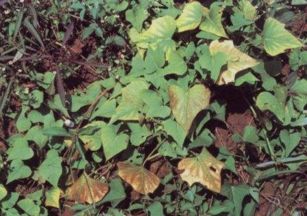
A Mg-deficient crop showing interveinal chlorosis and bronze-red
pigmentation on oldest leaves (J. Low).
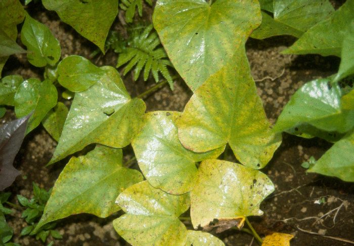
Typically the whole crop looks pale, and older leaves develop yellow
bands between main veins (P. Blamey).
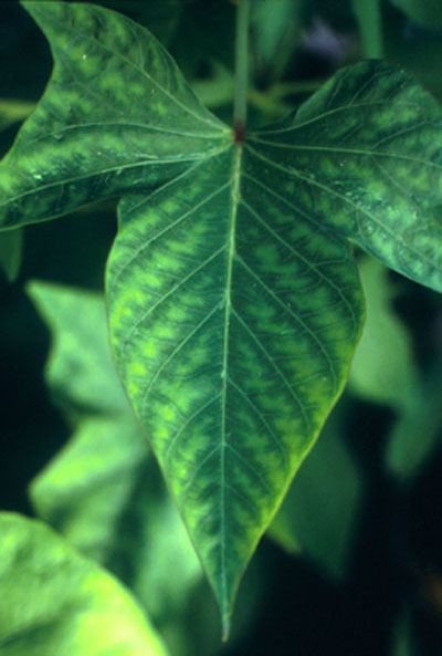
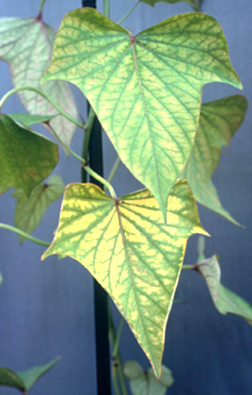
Interveinal chlorosis, with a margin of green either side of main veins,
progresses to yellowing (J.
O'Sullivan).
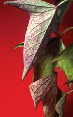
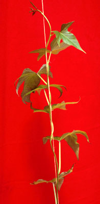
Symptoms sometimes
observed are purpling of veins under older leaves, and thin, elongated
twining growth of vine tips (J. O'Sullivan).
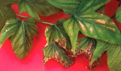
If necrosis develops before leaves wilt and fall, there are light
brown lesions spreading in interveinal and marginal chlorotic areas (J. O'Sullivan). |

